DPA/PICTURE ALLIANCE VIA GETTY IMAGES
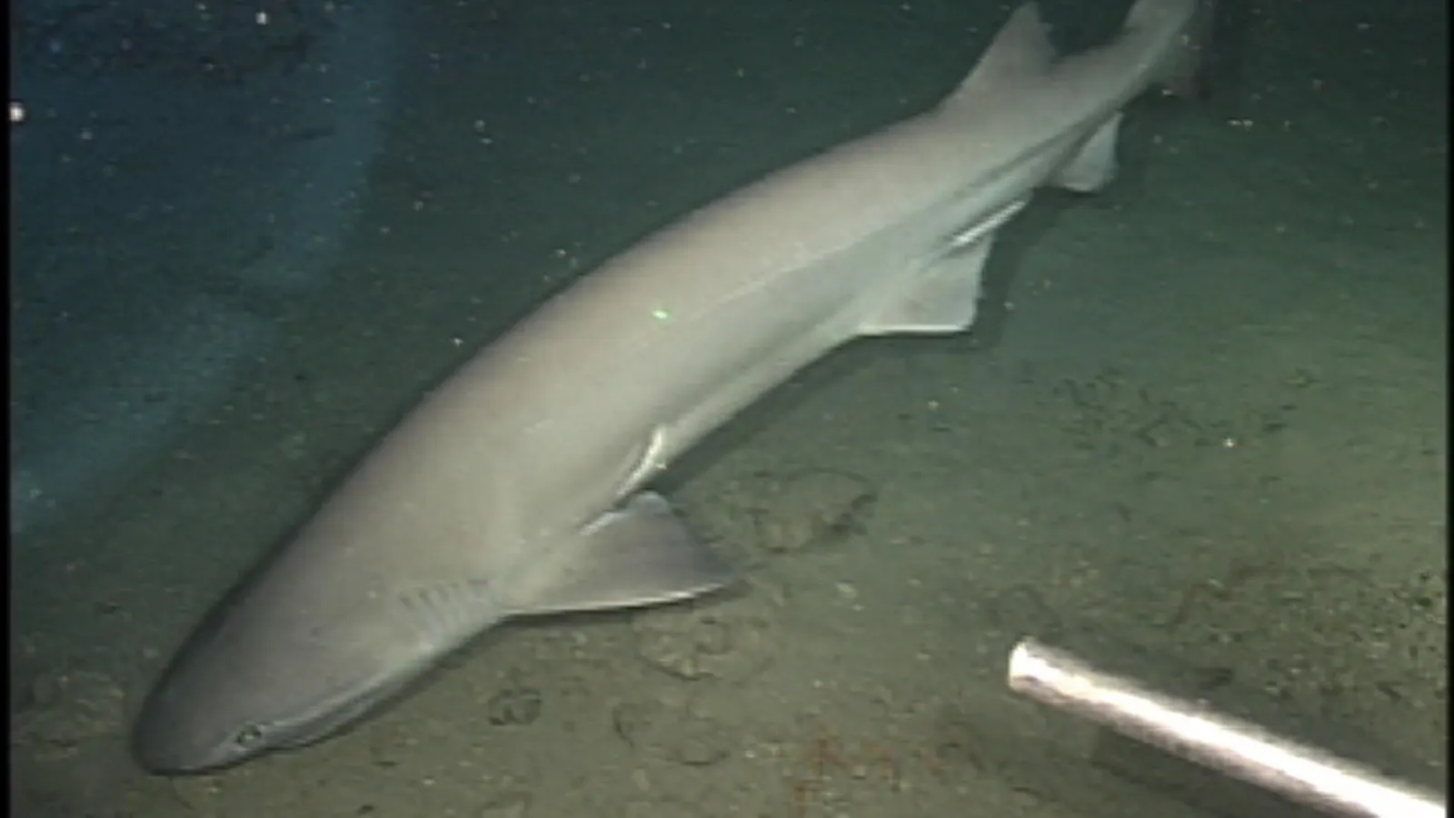
There is something deeply strange about looking at a shark embryo.
When you first look at it, it does not resemble the sleek predator most people imagine slicing through our ocean. Instead, it looks delicate, fragile and almost a bit alien. Tiny bulging eyes form long before the animal resembles a shark at all and its future face exists only as clusters of migrating cells, slowly organizing themselves into the structures that will eventually become jaws, cartilage and sensory organs. Yet hidden within that developing embryo is a story that stretches back more than 400 million years.
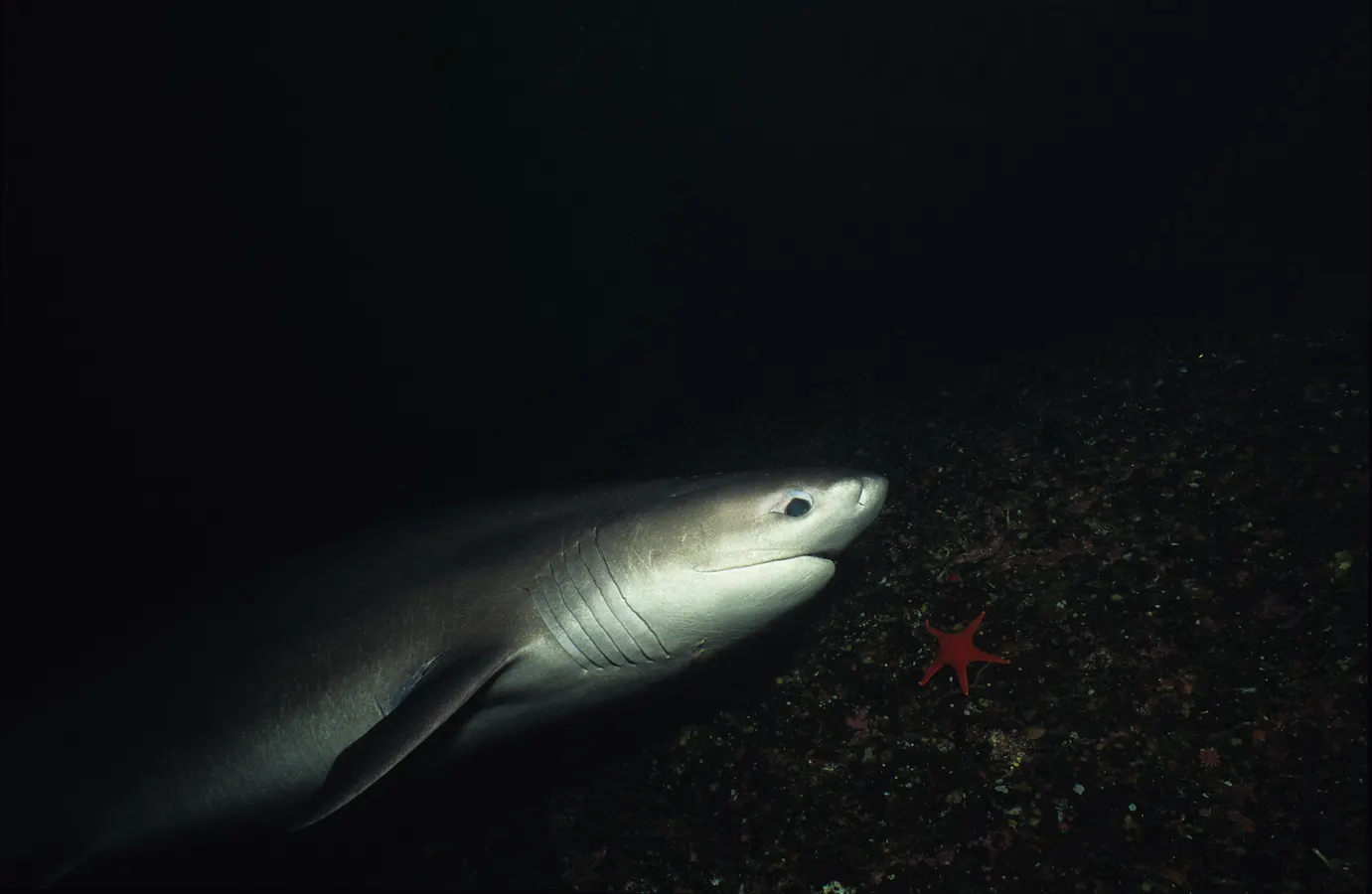
A new study led by Markéta Kaucká at the Max Planck Institute for Evolutionary Biology focused on the small-spotted catshark (Scyliorhinus canicula) is helping scientists better understand one of evolution’s most important cellular populations known as “neural crest cells.” These cells are unique to vertebrates and are often described as evolutionary game changers because they helped enable some of the defining features of vertebrate animals, including jaws, facial skeletons and advanced sensory systems. They emerge very early during embryonic development before migrating through the body and transforming into many different tissues: some become pigment cells, others help form parts of the nervous system. Cranial neural crest cells, the subset examined in this study, are especially important because they create much of the facial skeleton.