Skip to content
Unlike almost every other kind of microscope, atomic-force microscopes (AFMs) don’t use any kind of optical beam to image their subjects. Instead, they physically detect the subject’s surface with a tiny probe, repeating this thousands of times to build up a height map of the subject, sometimes with a resolution below a single nanometer. [Ben Krasnow] got to use an AFM in an investigation of one of his projects, and shared some unusual uses of it in his latest video.
For his first demonstration, [Ben] took a video of the probe head in action. Since the probe oscillates at nine kilohertz, this was less straightforward than it sounds, but a stroboscopic welding camera filming near that frequency could visualize its motion. The next project was to image some biological samples, particularly bacteria. First, [Ben] let the bacteria from nattō (fermented soybeans) multiply in a sterile growth medium, then centrifuged and washed them.
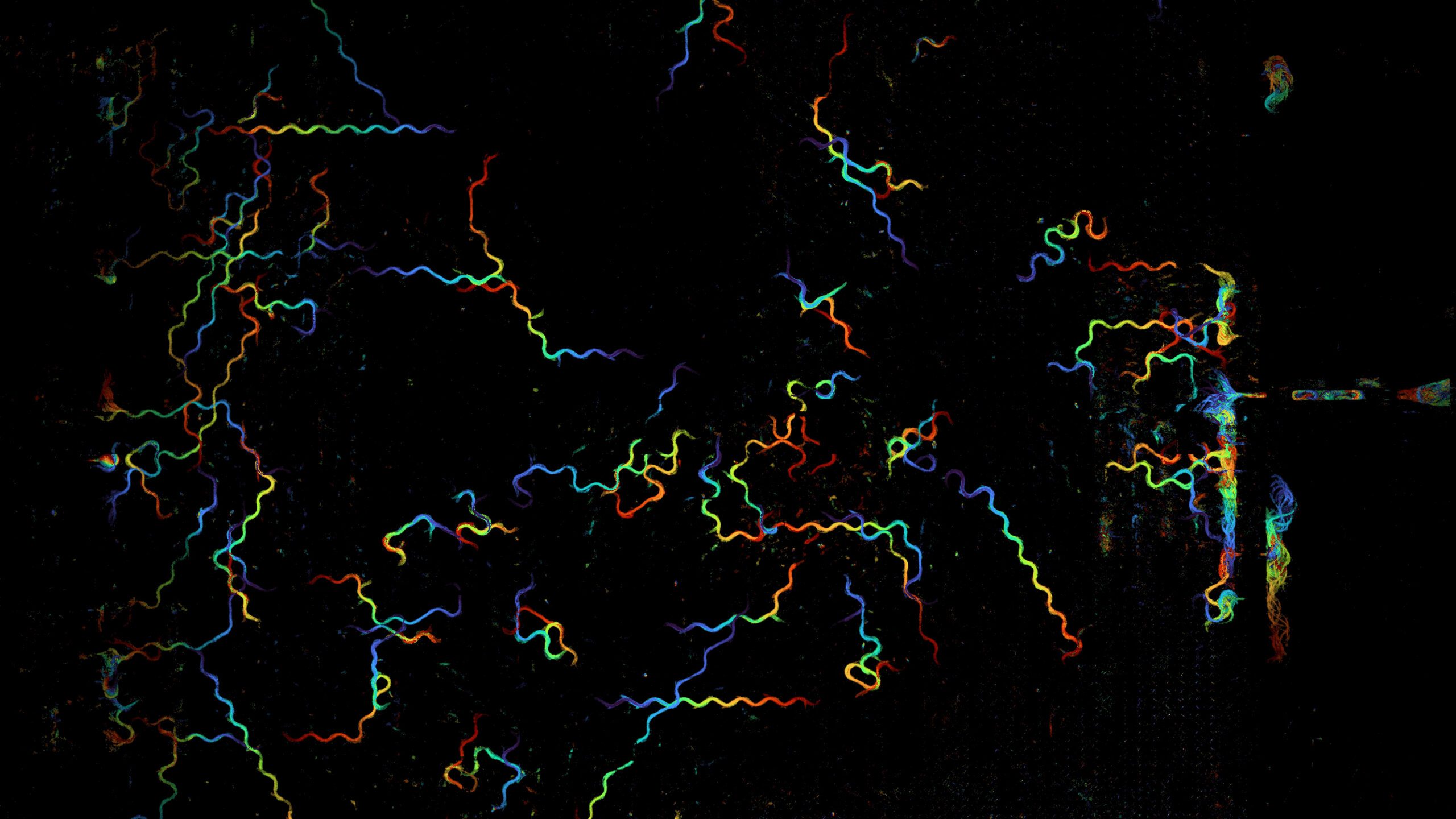
He spin-coated a thin layer of gelatine onto part of a silicon wafer, which provided a very flat substrate. The gelatine is electrostatically attracted to the bacteria, adhering them to the slide and letting [Ben] wash away other contaminants. This let the AFM image the bacteria clearly, even revealing how a spin-coating step had oriented them all in the same direction.