Nearly 100 years ago, a seemingly simple discovery revolutionized the microscope. The introduction of phase-contrast, which garnered a Nobel Prize in 1953, brought into clear view structures inside cells that had previously been too faint or washed out for biologists to study.
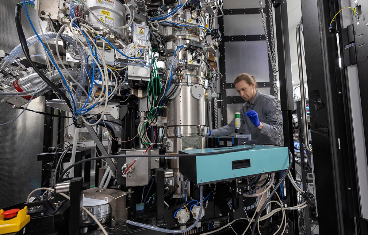
UC Berkeley physicists have now adapted the phase contrast technique to the electron microscope, which has about 10,000 times the magnification of microscopes using optical light.
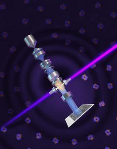
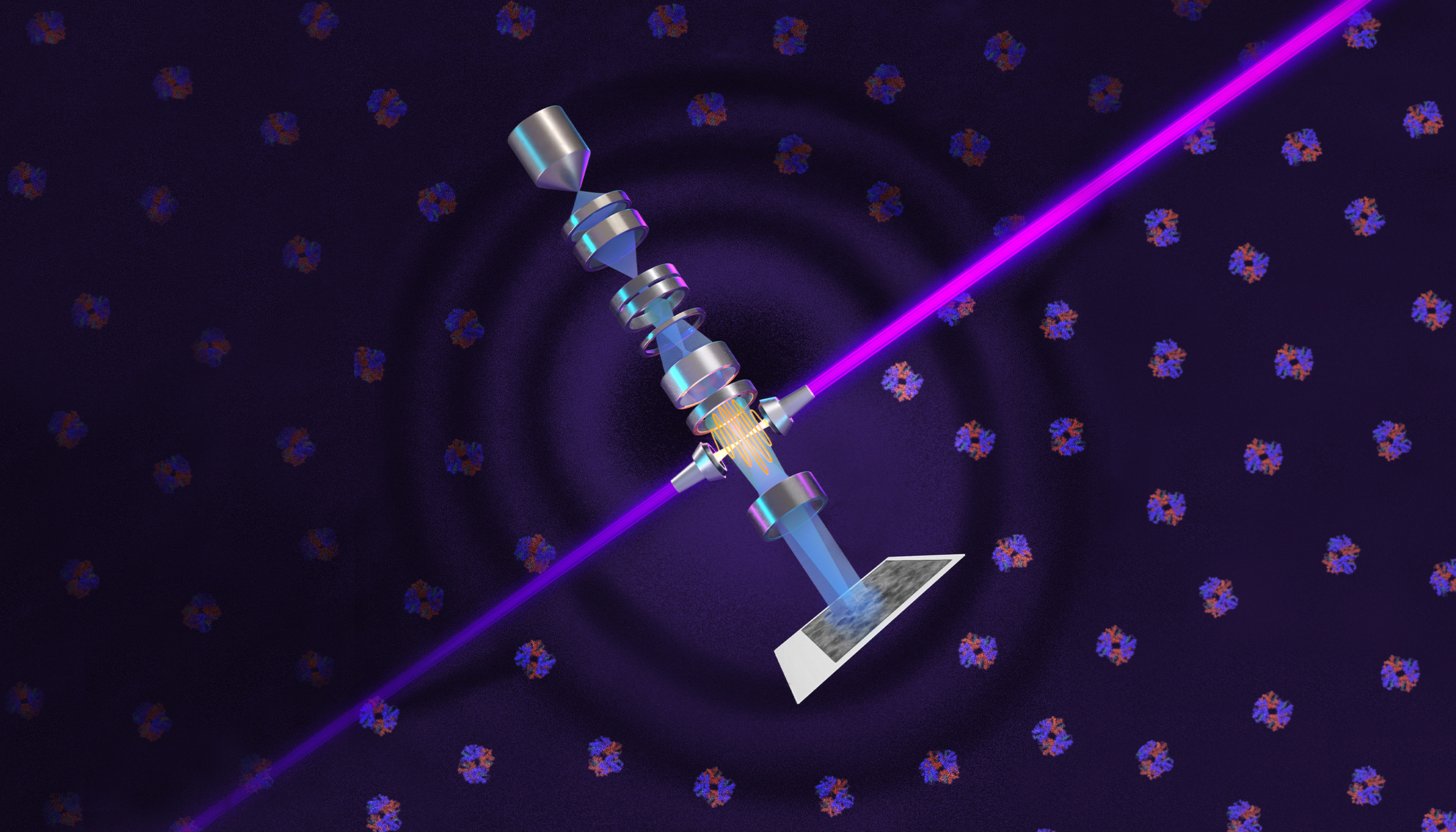
The addition of a so-called laser phase plate has the potential to greatly improve cryoelectron microscopy (cryo-EM), a technique for determining the structure of molecules that itself revolutionized the understanding of proteins and accelerated new drug discovery starting a decade ago. Despite its impact, however, cryo-EM still struggles to produce clear images of small molecules — including most human proteins. A laser phase plate promises clear images of most proteins in the cell down to one-third the size of those that are a challenge for today’s machines.
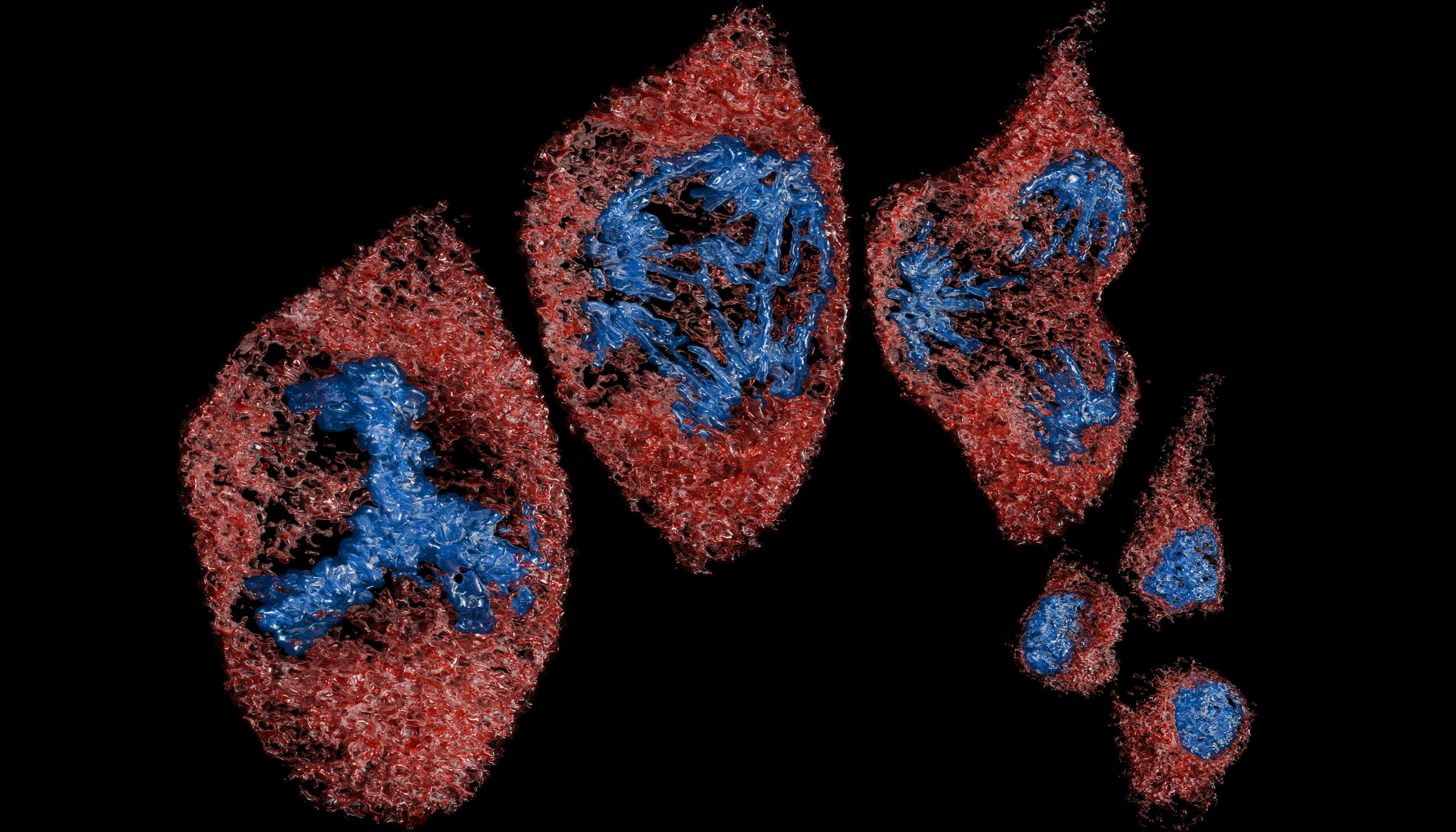
The addition of a laser phase plate seems certain to revolutionize a newer technique referred to as cryoelectron tomography (cryo-ET), which assembles a number of different angular views of a molecule or protein into a 3D image. This makes it possible to analyze proteins in their natural environment — inside cells — instead of in isolation in a solution.