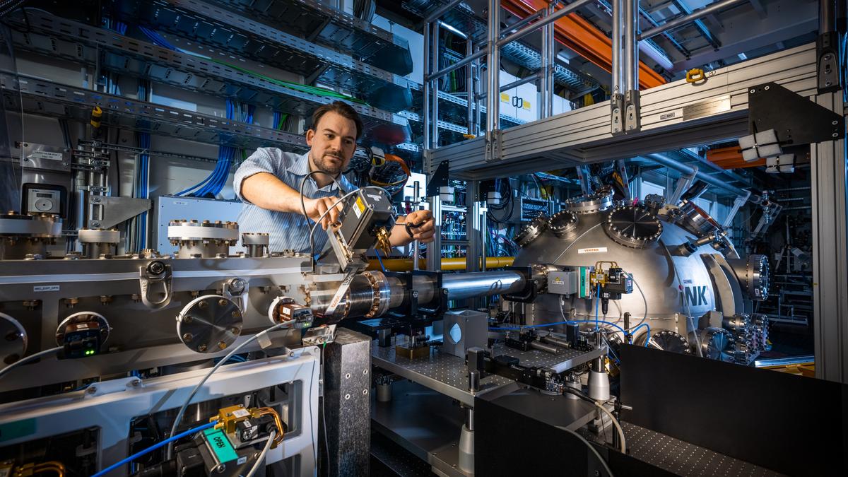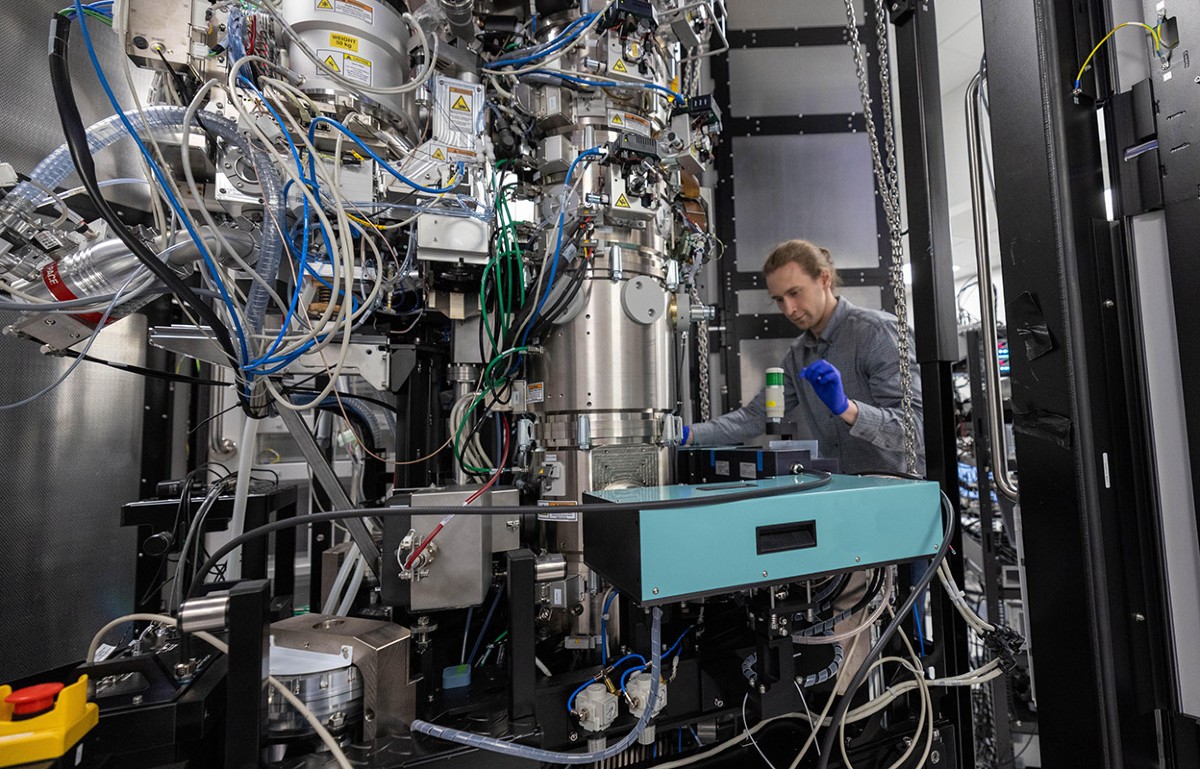
An engineer at Biohub in Redwood City, California, makes adjustments to the laser phase plate cavity inside a cryo-electron microscope.Credit: BiohubFor 15 years, structural biologists have debated the feasibility of a technology that could improve the resolution of protein structures produced by cryo-electron microscopy (cryo-EM). The technology, called a laser phase plate (LPP), should extend the cryo-EM technique to a broader range of proteins than was previously possible, and simplify tomography experiments that reconstruct protein behaviour in the cellular environment.This month, the optimists were vindicated, with a publication on 11 June in Science1 and a preprint posted in bioRxiv on 5 June2 demonstrating two LPP designs that effectively boost the image quality of small proteins in cryo-EM experiments.“It’s the first exciting new thing to happen in cryo-EM hardware beyond things that have been around for decades now,” says David Agard, founding scientific director of imaging at the biotechnology organization Biohub in Redwood City, California, who is an author of the preprint.‘This will never work’Cryo-EM involves freezing proteins in a thin layer of glassy ice and imaging them with an electron microscope. Biological materials generally do not absorb electrons, which makes them difficult to image directly. But they do scatter them, and this scattering alters a property of the electron wave known as the phase. Researchers have developed strategies to detect the resulting ‘phase shift’ by boosting the ‘phase contrast’, thereby enabling clearer protein imaging. In 2010, biophysicist Robert Glaeser and physicist Holger Müller at the University of California, Berkeley, teamed up to explore the idea of focusing an intense laser onto the electron beam used for cryo-EM imaging to selectively alter the phase of non-scattered ‘background’ electrons3. This would make it possible to increase the phase contrast of these electrons relative to those that have been scattered after encountering proteins.Cryo-electron microscopy wins chemistry NobelThey published a relatively rudimentary proof-of-concept prototype in 20194. This work caught the attention of Biohub, which has funded subsequent development. Müller recalls attending a meeting of cryo-EM specialists, hosted by the organization during the early days of the project, where he encountered considerable enthusiasm — but also deep scepticism. “The sentiment was, ‘This would be so cool, but this will never work,’” he says.Indeed, LPP development posed numerous technical challenges. “The laser focus that we have to generate is the brightest continuous laser focus anywhere,” says Müller. “And lasers of the required intensity didn’t exist when we started.” As a solution, he and his colleagues used precisely machined mirrors to reflect an incoming laser back and forth thousands of times, thereby amplifying it to the necessary intensity. These mirrors must be incredibly smooth — with a surface roughness of less than the diameter of a hydrogen atom — and are made of a ceramic glass that can resist damage from the intense laser beam, with further design features to ensure long-term stability.
An innovative technology boosts image quality for protein structures
After years of effort, two research teams have developed ‘laser phase plate’ systems that could help cryo-electron-microscopy users to generate high-quality structures for a broad range of proteins.
TL;DRAI
Müller and Glaeser's laser phase plate (LPP), published in Science, resolves 15-year technical debate to improve small protein cryo-EM imaging. First cryo-EM hardware advance in decades; extends protein range and streamlines tomography for biotech and pharma structural discovery.
476 words~2 min read