view more
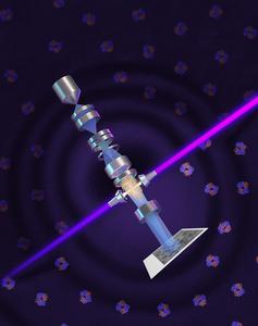
A team led by Raju Tomer, professor of biological sciences at Columbia University, has created a new design for microscopes and microscope lenses that could push 3D tissue imaging beyond state-of-the-art systems while drastically cutting costs and complexity. Details of the design were published today in the journal Nature Biotechnology.
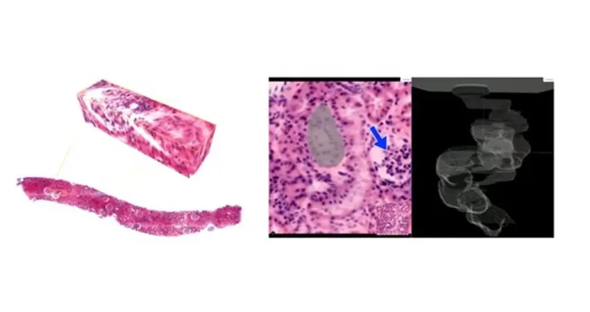
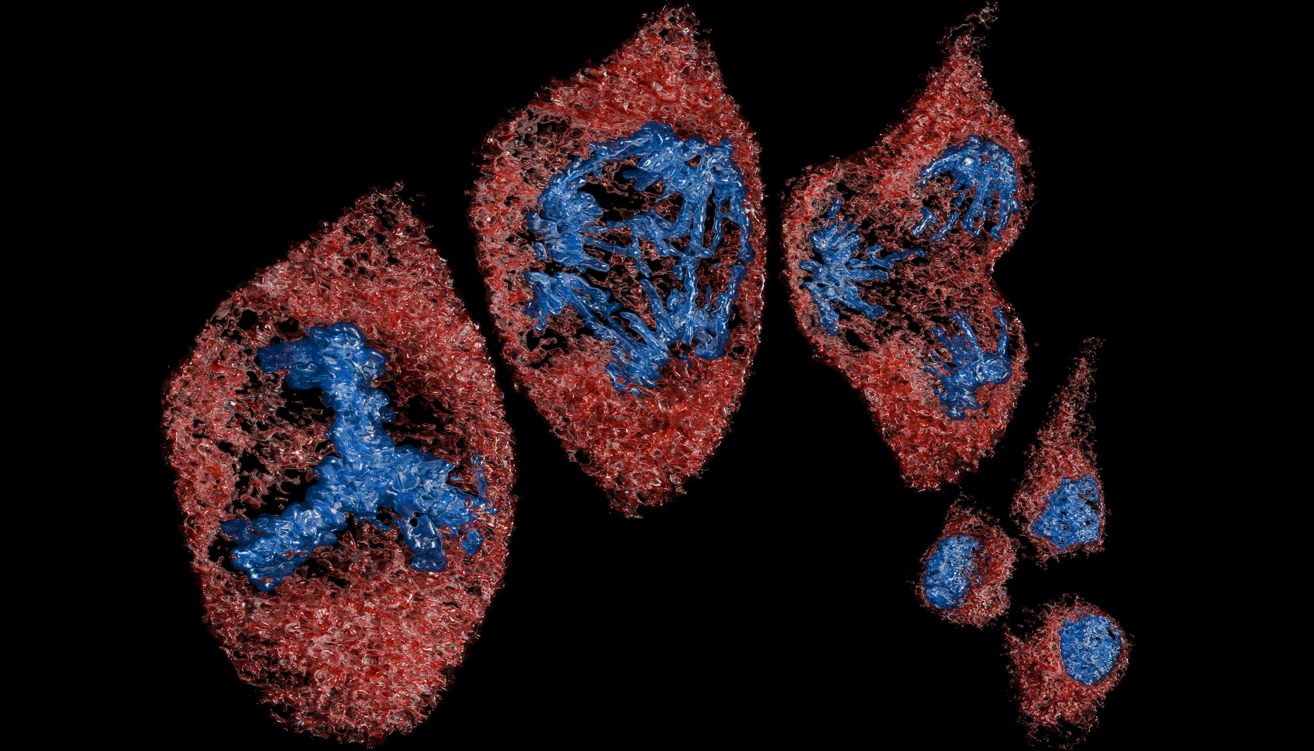
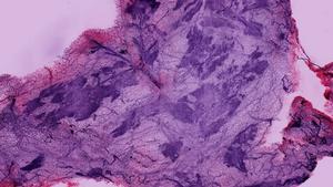
Modern biology and medicine increasingly depend on high-resolution, 3D images of intact tissues such as brains and cancer biopsies. The images allow researchers to map neural circuits, characterize disease, and train next-generation AI models for diagnosis, among other uses. But progress in imaging these tissues easily and at a large scale has been bottlenecked by the lenses that capture light from samples. Researchers have had to make tradeoffs: “Oil-immersion” lenses, which touch the sample through a drop of oil, deliver the sharpest images; but they’re expensive, can only see a few millimeters deep at most, and require specific sample preparations. Cheaper lenses that work at a distance through air can reach up to several centimeters into a sample, but they produce blurred images when used with the chemicals that render tissues transparent for 3D viewing.