By advancing three-dimensional imaging of tissue at the microscopic scale, tissue-based analysis of changes in gene expression, and artificial intelligence (AI) methodologies, the team aims to generate new insights from tissue samples into health and disease. This £5M programme is made possible through funding from the Wellcome Trust Bioimaging Technology Development initiative.
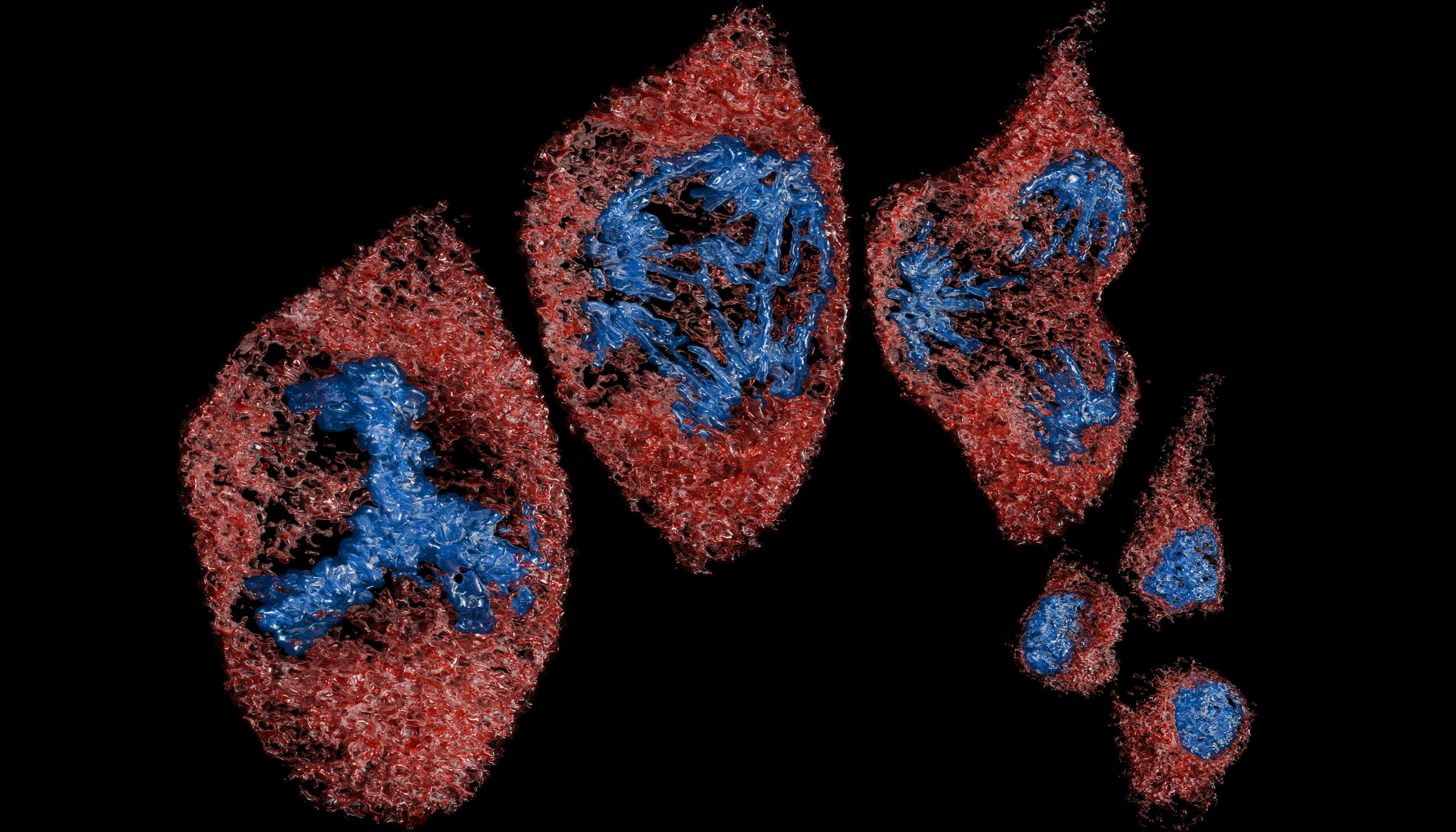
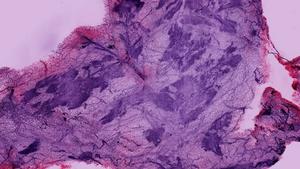
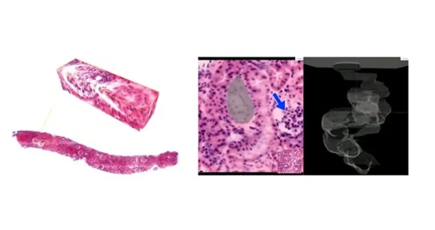
Today, pathologists diagnose disease by analysing razor thin two-dimensional (2D) tissue sections under a microscope. But all components of the tissue, as for example cells and blood vessels, are three-dimensional (3D) objects. This is why the team will investigate how the analysis of the complex 3D tissue anatomy can aid the development of more informative disease models. Work in prostate cancer, bone marrow and skeletal health, and kidney disease, aims to connect fundamental pathological processes such as the build up of scar tissue (fibrosis), inflammation, and changes in the vasculature. Using a concrete set of studies, the research programme will demonstrate potential impact on human health.
The programme is led by Professor Jens Rittscher, and involves medical image analysis specialist Professor Konstantinos Kamnitsas, kidney disease expert Katherine Bull (Nuffield Department of Medicine), prostate cancer scientist Ian Mills (Department of Surgical Sciences), and bone marrow expert Daniel Royston (Radcliffe Department of Medicine). In Cape Town, Bianca Davidson leads the kidney disease study with clinical scientist Nicola Wearne (Groote Schuur Hospital), pathologist Brendon Price (Anatomical Pathology), and imaging expert Michael Reiche (Institute of Infectious Disease and Molecular Medicine). Internationally, the team will collaborate with Yale University, Charite in Berlin and US National Cancer Institute.