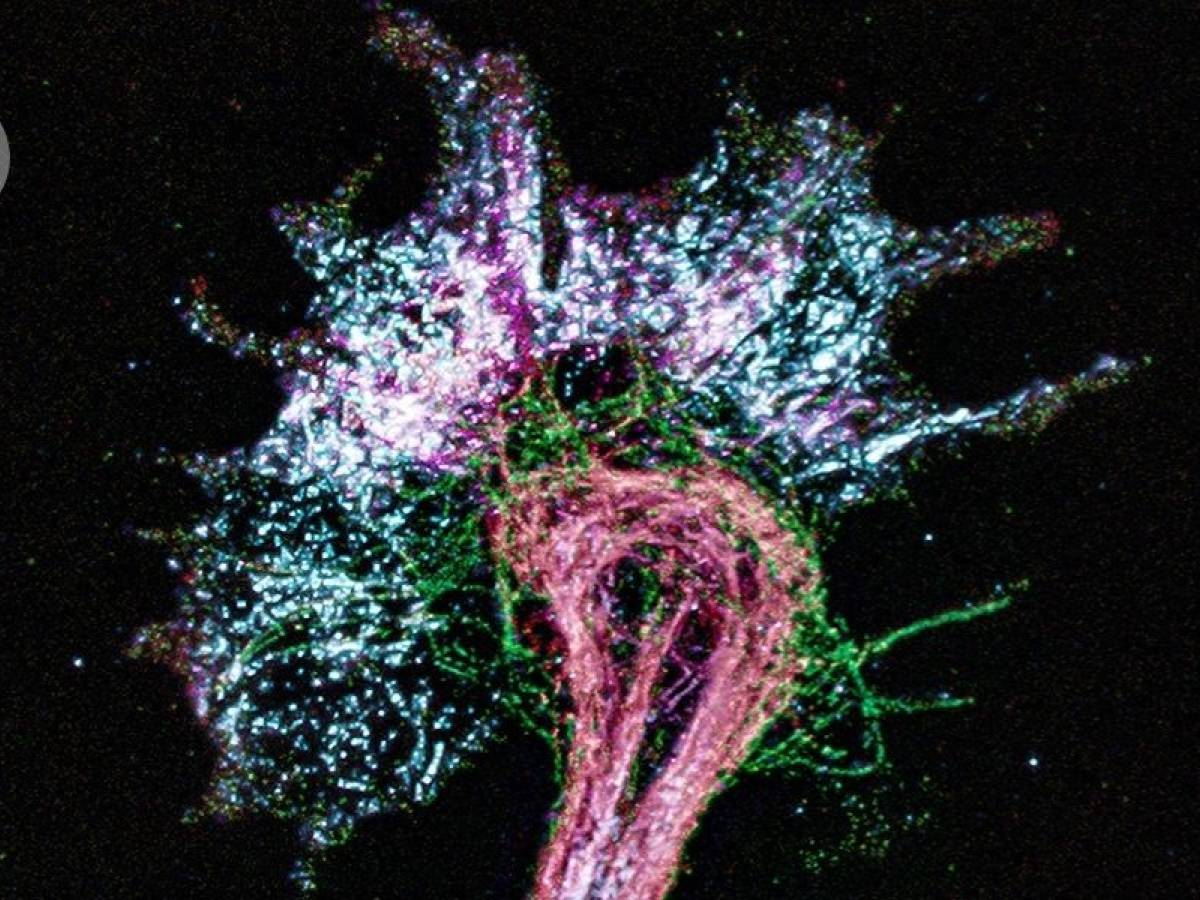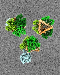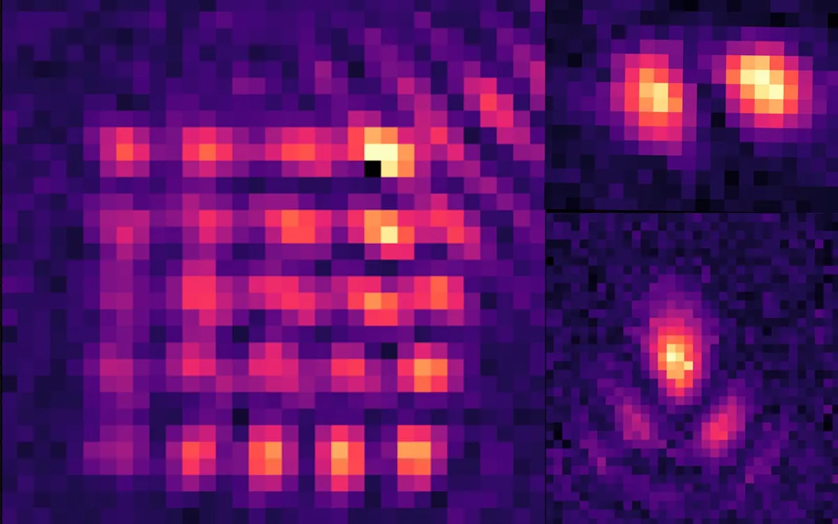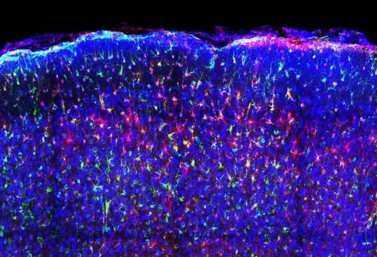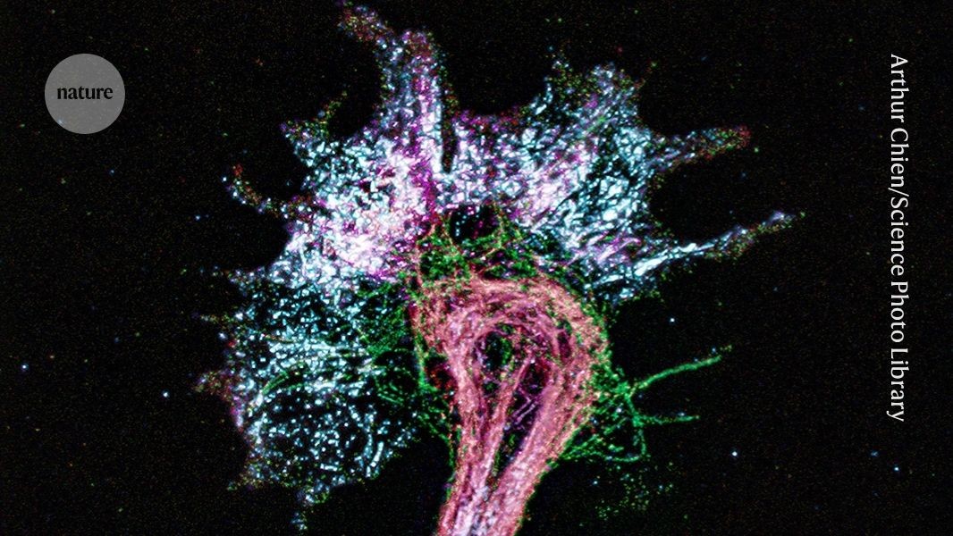
High-resolution images can be produced by increasing the size of a nerve cell before fluorescence-microscopy imaging.Credit: Arthur Chien/SPLA technique that supersizes cells to reveal minute details has gone big — really big.Using a polymer like those used in nappies to make them super-absorbent, scientists have expanded the volume of biological samples so they are one billion times bigger — 1,000-fold larger in each dimension. This level of expansion could inflate an individual cell to the size of a mouse brain and a US dime-size sample to the proportion of an Olympic swimming pool.Researchers have used the technique to map the positions of amino acids (the building blocks of proteins) within proteins and small molecules called peptides using conventional light microscopes. The approach is outlined in a preprint posted on bioRxiv earlier this month1.Previously, viewing molecules in such fine detail has typically been achieved by using costly and complicated techniques such as cryogenic electron microscopy (cryo-EM) and X-ray crystallography. “This is the democratization of structural biology,” says study co-author Silvio Rizzoli, a neuroscientist and imaging specialist at the University Medical Center Göttingen (UMG) in Germany.“It’s really getting down to a ground-truth description of what a protein is,” adds co-author Helena Hu, a bioengineer at the Massachusetts Institute of Technology (MIT) in Cambridge.Expansion microscopyThe laws of physics limit the optical power of light microscopes: objects that are less than about 200 nanometres apart cannot be distinguished. ‘Super-resolution’ light microscopy techniques, which usually rely on optical tricks and costly kit, have cut this resolution limit to below 10 nm.In 2015, MIT neuroengineer Edward Boyden, a co-author on the study, and his colleagues invented an alternative way to get extremely high resolution using ordinary fluorescent light microscopes2. Using a swelling hydrogel, the researchers expanded tissue samples about fourfold in each direction. This moved cellular components away from one another, improving resolution.Researchers have since adapted Boyden’s 'expansion microscopy' method to swell samples to greater volumes, but inflation has generally been limited to about a 20-fold increase in size.To go bigger, the researchers developed a new hydrogel recipe in which samples were expanded multiple times. The team also took an expansion-microscopy approach called ONE microscopy3, to map protein structures with a light microscope. Purified proteins were bonded to the hydrogel and then broken apart using enzymes or heat, enabling proteins to be stretched apart without compromising their 3D shape.Gigantic proteinsUsing their 1,000-fold expansion method, the researchers labelled several of the nine individual amino acids that make up a peptide called mCLING. They then imaged the relative locations of the building blocks using a fluorescent microscope. These measurements were consistent with structures of mCLING derived from computational simulations.The method also revealed the structure of a protein called a nanobody (a small version of an antibody), as well as the locations of some of its amino acids. Furthermore, the researchers used the technique to map the previously determined structure of green fluorescent protein (GFP), a biotechnology workhorse used to label other proteins.Expansion microscopy opens the door to exploring more challengesThe researchers estimate that their model of GFP — reconstructed from thousands of snapshots — attained a resolution of around 1.2 nm, or 12 ångstroms. This is far from a 1.9-Å structure of GFP available in a public repository and determined using X-ray crystallography, or the level of detail that scientists routinely achieve using cryo-EM.But co-author Ali Shaib, a nanoscale specialist at UMG, says that methodological improvements have driven resolution below 10 Å for purified proteins, in unpublished work. Rizzoli notes that the team is now able to determine the shapes of proteins that are still inside cells at around 10 Å. "We are trying to make cryo-EM like structures from affordable optical microscopes," Shaib says.
Making samples one billion times bigger lets simple microscopes pinpoint amino acids
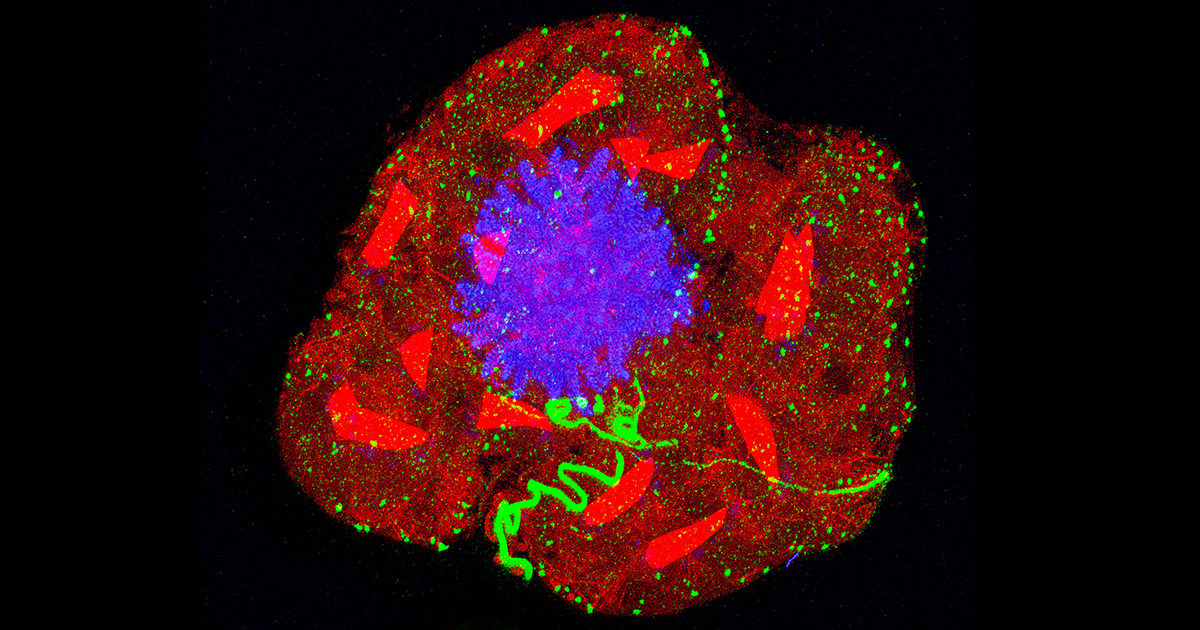
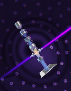
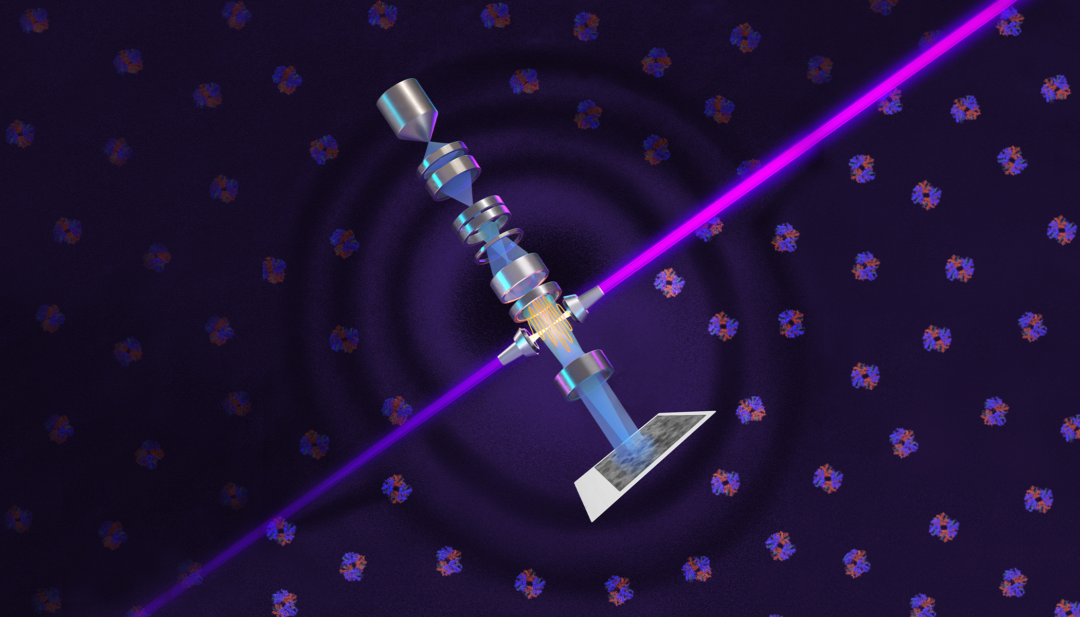
Stretching protein samples in all directions pulls molecules farther apart, allowing them to be visualized using only light microscopy.
617 words~3 min read