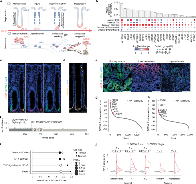
MainThe intestinal epithelium is characterized by a hierarchy of cell states that balance self-renewal and differentiation to maintain homeostasis2. Crypt base ISCs, which are marked by the expression of genes such as LGR5, drive the continuous production of short-lived, specialized differentiated daughter cells that are eventually shed into the intestinal lumen16. However, when LGR5+ ISCs are lost during injury, the epithelium displays substantial plasticity. A broad range of differentiated cells have the capacity to undergo injury-induced reprogramming. Initially, cells enter into a transient regenerative state with fetal-like properties and then they dedifferentiate into a LGR5+ ISC state to repopulate the crypt base and restore homeostasis2,3,17 (Fig. 1a). Although the transcription factor ASCL2 is necessary for this process18, little is known about injury-induced triggers of dedifferentiation. In CRC, the capacity of the intestinal epithelium to undergo stress-induced reprogramming contributes to tumour progression, metastasis and therapy resistance19. Primary tumours largely adopt LGR5+ ISC states either through the acquisition of oncogenic driver mutations in crypt base ISCs or through the dedifferentiation of mutated differentiated cells into tumour-permissive ISC-like states20,21,22,23. Cells at the primary tumour-invasion front that detach from the tumour mass enter into LGR5– metastasis-initiating states marked by increased expression of injury-associated regenerative and fetal markers (for example, L1CAM, PROX1, TROP2 and ANXA1)7,10,24. These cells also exhibit increased expression of epithelial differentiation markers (for example, EMP1)9, and are associated with increased activity of the AP-1 family of stress-response transcription factors25,26 (henceforth termed an ‘injury–repair’ state). After extravasation in distant organs, differentiated LGR5– metastasis-initiating cells can dedifferentiate into canonical LGR5+ ISC states to seed metastasis and regenerate tumours, a process that mirrors the restoration of homeostasis during intestinal regeneration8,9,10,26,27,28 (Fig. 1a). Alternatively, CRC metastases can undergo lineage plasticity into non-canonical squamous-like and neuroendocrine-like states not observed during regeneration in the intestinal niche10,26. Such non-canonical states are common among patients and are associated with adverse clinical outcomes compared with patients with tumours that primarily express canonical states. The mechanisms that underpin such cell-state reprogramming and cell-fate decision-making are largely unknown. To gain insight, we sought to identify the molecular switch that controls stress-induced dedifferentiation into a LGR5+ ISC state, a conserved process that is essential for both epithelial regeneration and canonical metastases.Fig. 1: ZFP36L2 is expressed by ISCs and is associated with the AP-1 gene program.a, Schematic of cell-state plasticity and dynamics during intestinal regeneration (top) and CRC (bottom). b, Dot plot of tumour ISC-like module10 gene expression in normal colon (Normal) and CRC (Tumour) epithelial cells from 25 patients. The dot size indicates the per cent cells (rows) expressing each gene (columns). Colour scale indicates the log2 fold change in average gene expression. Bar plots show the mean expression of each gene across normal colon ISCs. Genes meeting false discovery rate (FDR) < 0.05 using the Wilcoxon rank-sum test (ISC versus all other epithelial subtypes) are shown. c,d, Representative LGR5 and ZFP36L2 RNA FISH (c) and ZFP36L2 IF (d) images of human normal colon crypts. e, Representative LGR5 and ZFP36L2 RNA FISH images of human primary CRC tumours (n = 2), liver metastases (n = 2) and lung metastases (n = 2) from 2 donors. f, Lollipop plot showing ZFP36L2 mutation frequency in the TCGA63, DFCI64 and MSKCC whole-exome sequencing CRC cohorts. n = 96 tumours. g,h, ZFP36L2 autocorrelated gene programs in normal colon (g) and CRC epithelial cells (h). Red, top AP-1 family genes65. n = 12,955 normal colon (24 donors) and 25,317 CRC cells (25 patients). FDR < 0.05. i, GSEA of genes (Supplementary Table 1b–f) ranked by positive ZFP36L2 autocorrelation (Methods), which revealed conserved pathways enriched in normal (white dots) and tumour (black dots) epithelial cells. n = 12,955 normal colon (24 donors) and 25,317 CRC cells (25 patients). FDR < 0.05. j, Violin plots showing AP-1 pathway gene expression in ZFP36L2-high and ZFP36L2-low populations (top and bottom 10 percentile expression). n = 1,806 differentiated, n = 1,326 TA and n = 230 ISC cells from normal colons (24 donors) and 2,622 primary CRC and 2,444 CRC metastasis cells (25 patients). Two-sided Mann–Whitney U-test. Scale bars, 50 µm (c,d) or 200 µm (e).ZFP36L2 co-expression with stress programsTo identify mediators of cell-state switching, we focused on genes co-expressed in ISC-like cells in a cohort of same-patient trios of normal, primary and metastatic CRC tissue samples. Genes were identified through Hotspot gene module analyses of single-cell RNA sequencing (scRNA-seq) data from fresh tissue10, and we termed them ‘tumour ISC-like’. ZFP36L2 (also known as TIS11D, ERF2 and BRF2) stood out as the gene most highly expressed in ISC-like cells. ZFP36L2 expression progressively decreased with differentiation in both normal intestinal crypts and in CRC, including canonical intestinal and non-canonical lineages (Fig. 1b–e and Extended Data Fig. 1a–d). Whole-exome and whole-genome sequencing studies11,12,13,14,15 have identified recurrent ZFP36L2 mutations in 5–10% of tumours from patients with CRC (Extended Data Fig. 1e,f). Most of these mutations are frameshift deletions with truncation of the coding region upstream of the zinc finger domain, which probably results in loss of ZFP36L2 function (Fig. 1f). ZFP36L2 is an RNA-binding protein of the ZFP36 (also known as tristetraprolin) family of CCCH-tandem zinc finger proteins. These proteins bind AU-rich elements in 3′ untranslated regions (UTRs) and recruit the RNA exosome or the CCR4–NOT complex to mediate mRNA degradation in haematopoiesis and early embryogenesis29,30,31,32. However, little is known about ZFP36L2 function in epithelial cells and cancer.To gain insight, we performed gene expression autocorrelation analysis across normal ISCs, transit-amplifying (TA) cells and tumour cells (Methods). Genes most strongly co-expressed with ZFP36L2 in both normal colon and CRC samples included AP-1 transcription factors and other components of the AP-1 stress-response pathway (for example, FOSB, FOS, MYC, EGR1, MYB, ATF3 and DUSP1; Fig. 1g–j, Extended Data Fig. 1g and Supplementary Table 1c–f). Although ZFP36L2 was most broadly expressed in tumour ISC-like cells, a subset of ZFP36L2-high CRC cells displayed an injury–repair state marked by high expression of the RevCSC signature28, and the metastasis-initiating cell markers L1CAM7 and EMP19 (Extended Data Fig. 2). The AP-1 transcription factor complex, formed by FOS, JUN and ATF family of heterodimers, has a central role in orchestrating stress-induced transcriptional responses during tissue regeneration33. AP-1 is rapidly activated in response to cytokines, growth factors or microbial or physical stressors. Subsequently, AP-1 integrates upstream signals—primarily via MAPK, JNK and ERK pathways—to drive expression programs that regulate proliferation, survival, differentiation and epithelial remodelling. ZFP36 family members are well-characterized immediate-early genes and are rapidly induced in response to such stimuli34,35,36,37. EGF stimulation of cells leads to the sequential induction of AP-1 followed by ZFP36L235, which indicates that ZFP36L2 has a role downstream of mitogenic signalling. Together, these data suggest that ZFP36L2 might be a molecular link between AP-1-driven stress response in the injury–repair state and re-entry into the ISC state.ZFP36L2 mediates dedifferentiationWe first explored the relationship between ZFP36L2, the stress response and the ISC state. As in humans, mouse Zfp36l2 mRNA (Fig. 2a) and ZFP36L2 protein (Fig. 2b) were most highly expressed in crypt base ISCs, with expression declining with differentiation. Colitis induced by dextran sodium sulfate (DSS) leads to destruction of intestinal epithelial cells (IECs), followed by dedifferentiation of surviving differentiated progenitors into LGR5+ ISCs and restoration of homeostasis once DSS is withdrawn38. ZFP36L2 expression increased by day 2 of DSS administration, concurrent with LGR5 loss, whereas both ZFP36L2 and LGR5 expression were downregulated at the peak of injury by day 7 (Fig. 2c). During regeneration, ZFP36L2 was re-expressed first in the mid-crypt (TA compartment) and crypt base (ISC compartment) by day 10, preceding restoration of crypt base Lgr5 expression by day 14.Fig. 2: Zfp36l2 is required for injury-induced dedifferentiation during intestinal regeneration.a, Zfp36l2 expression across epithelial cells, as measured by scRNA-seq of colonic crypts (21,000 cells from 11 mice)66. Marker genes for each cell type are indicated by boxes. b, Representative images (left) and quantification (right) of Lgr5 RNA FISH and ZFP36L2 and MUC2 IF in colon tissue from 7-week-old C57BL/6J mice based on the cell position along the crypt base (position 0) to lumen axis. n = 2,275 cells from 31 crypts. c, Representative images of Lgr5 RNA FISH and ZFP36L2 and MUC2 IF in colon tissue collected at the indicated time points from mice treated with 3.5% DSS for 7 days followed by recovery for 7 days. n = 3 mice per time point. Expanded fields of view are provided in Supplementary Figs. 1 and 3. d, Top, schematic of the experiment. Bottom, daily body weight measurements. n = 5 (water) mice; n = 10 (wild-type (WT)) and 14 (IEC) (DSS-treated) mice. Mean (solid line; dots, individual time points) ± s.e.m. (shaded area; dashed lines, s.e.m. boundaries); two-tailed Mann–Whitney U-tests at day 7 and day 14. e, Caecum (top) and colon (bottom) lengths on day 14. n = 3 mice (water); n = 10 (WT) and 12 (IEC) (DSS-treated) mice. Mean ± s.d.; two-tailed Mann–Whitney U-test. NS, not significant. f, Lgr5DTR ablation. Top, schematic of the experiment. Bottom, representative images (n = 2 mice) of colon sections stained by Lgr5 RNA FISH or ZFP36L2 IF at the indicated time points. Expanded fields of view are provided in Supplementary Fig. 5. Controls: Lgr5DTR-negative mice treated with 4 doses of DT over 7 days. g,h, Mice (8–10 weeks old) were administered DT every 2 days for 4 doses. Mice were euthanized and colons were collected 12 days after the first DT dose. g, Images of mouse Lgr5 and Zfp36l2 mRNA FISH on day-12 Swiss-roll colon sections. h, The number of mice showing regeneration of the Lgr5 FISH signal. i, Schematic of the expression and function of ZFP36L2 in injury-induced dedifferentiation to drive regeneration in the mouse colon. Scale bars, 50 µm (b,c,f,g).Source dataThese observations of a temporospatial sequence of ZFP36L2 and LGR5 expression during crypt regeneration suggested that ZFP36L2 might have a functional role in the injury-induced dedifferentiation of TA progenitors into a LGR5+ ISC state. To test this hypothesis, we generated Vil1creZfp36l2fl/fl mice to knockout Zfp36l2 in IECs39,40 (henceforth called Zfp36l2IEC mice) and Vil1creZfp36l2+/+ (Zfp36l2WT) and Vil1creZfp36l2fl/+ (Zfp36l2HET) littermate controls (Extended Data Fig. 3a,b). Zfp36l2IEC mice displayed normal colon morphology and body weight (Extended Data Fig. 3b-d). Immunofluorescence (IF) and RNA fluorescence in situ hybridization (FISH) staining of the colon revealed a reduction in LGR5+ ISCs from Zfp36l2IEC mice (Extended Data Fig. 3e,f). This result was quantified by crossing Zfp36l2IEC and Zfp36l2WT mice with Lgr5eGFP-creERT2 reporter mice16 and performing flow cytometry for LGR5–eGFP+ cells on isolated colon crypts (Extended Data Fig. 3g–i). Thus, at homeostatic baseline, Zfp36l2IEC mice seem to maintain normal intestinal morphology and function despite having a reduced LGR5+ ISC population. To determine the function of intestinal epithelial ZFP36L2 during stress, we treated mice with 3.5% DSS for 7 days to induce intestinal injury, followed by 7 days of regeneration once DSS treatment was stopped. Zfp36l2IEC mice displayed increased weight loss during DSS treatment and incomplete weight recovery 7 days after DSS withdrawal relative to Zfp36l2WT controls. This result was consistent with more substantial epithelial injury and impaired regeneration after Zfp36l2 knockout (Fig. 2d). Pathology revealed shortening of the colon and caecum with persistent inflammation, crypt loss and decreased Lgr5 expression at day 14 in Zfp36l2IEC mice compared with Zfp36l2WT controls (Fig. 2e and Extended Data Fig. 3j–m).To determine whether epithelial cell-intrinsic mechanisms inhibit dedifferentiation in colonic epithelia of Zfp36l2IEC mice, we used an orthogonal Lgr5DTR mouse model in which LGR5+ ISCs are directly ablated using diphtheria toxin (DT)5. Direct ablation of LGR5+ ISCs induced Zfp36l2 expression first in the TA compartment, with expression moving down the crypt and preceding the restoration of crypt base Lgr5 expression (Fig. 2f and Extended Data Fig. 4a). Next, we generated Lgr5DTR-eGFP+Zfp36l2IEC and Lgr5DTR-eGFP+Zfp36l2WT mice, administered DT to selectively ablate LGR5+ ISCs and assayed the restoration of Lgr5 expression on the basis of the dedifferentiation of LGR5– cells (Fig. 2g,h). Consistent with results from the DSS-induced colitis model, loss of Zfp36l2 impaired the recovery of LGR5+ crypt base ISCs following the direct ablation of LGR5+ cells. To exclude potential confounders from in vivo DT administration, we established organoids from the colon epithelium of Lgr5DTR-eGFP+Zfp36l2IEC and Lgr5DTR-eGFP+Zfp36l2WT mice. Organoids were treated with vehicle or DT at day 4 for 24 h, and DT-dependent ISC ablation was verified by flow cytometry for LGR5–eGFP expression (Extended Data Fig. 4b,c). Flow-sorted single cells were seeded at low density to facilitate the regeneration of new organoids, a function that requires dedifferentiation into an LGR5+ ISC state41. Lgr5DTR-eGFP+Zfp36l2IEC organoids regenerated significantly fewer organoids than Lgr5DTR-eGFP+Zfp36l2WT controls (Extended Data Fig. 4e,f). Flow cytometry confirmed re-expression of LGR5–eGFP, with reduced LGR5–eGFP re-expression in organoids recovered from Lgr5DTR-eGFP+Zfp36l2IEC mice (Extended Data Fig. 4d). Thus, Zfp36l2 deletion inhibits the dedifferentiation of LGR5– cells into LGR5+ cells and impairs organoid regeneration after DT-mediated LGR5+ ISC ablation.We also established colon organoids from Zfp36l2IEC, Zfp36l2HET and Zfp36l2WT mice by plating intact crypts (Extended Data Fig. 4g–l). After 3 days of ISC culture in mWRENAFI medium41 (Methods), WNT and R-spondin1 were withdrawn to induce differentiation for 3 days (Extended Data Fig. 4g–i). To assess dedifferentiation capacity, differentiated organoids were dissociated into single cells, flow-sorted for live cells and reseeded at low density to regenerate new organoids. Zfp36l2IEC cells re-established fewer and smaller organoids than Zfp36l2WT and Zfp36l2HET controls (Extended Data Fig. 4j–l). In summary, both the DSS and Lgr5DTR ablation models demonstrated that ZFP36L2 is required for injury-induced dedifferentiation of IECs into an ISC state, which in turn is required for epithelial regeneration and wound healing (Fig. 2i).Loss of ZFP36L2 abrogates CRC metastasisAs in tissue repair, disseminating metastasis-initiating cells enter injury-induced differentiated states. However, they can also dedifferentiate into an ISC state during metastatic seeding7,8,9,10 (Fig. 1a). To determine whether ZFP36L2 has a role in dedifferentiation into an ISC state during CRC progression, we engineered four patient-derived CRC organoid lines and used doxycycline-inducible short-hairpin RNAs (shRNAs) to knockdown ZFP36L2 (shZFP36L2). Scrambled shRNA was used as a control (shCtrl) (Extended Data Fig. 5a–d). We also sought to clarify how the underlying canonical or non-canonical differentiation potential of the tumour cell population influences ZFP36L2 function. Therefore, we included two primary tumour-derived organoids (MSK125P and OKG146P) with canonical gene expression (largely ISC with some intestinal differentiation)10 and two metastasis-derived organoids (MSK107Li and OKG146Li) with both canonical and non-canonical differentiation potential7,10 (Extended Data Fig. 5a,b). To assess the role of ZFP36L2 in tumour growth in the intestinal niche, organoids transduced with shZFP36L2 or shCtrl were orthotopically injected into the caecum of NSG mice, and animals were administered doxycycline to knockdown ZFP36L2 (Fig. 3a). Primary tumour-derived canonical organoids (OKG146P and MSK125P) exhibited decreased primary tumour growth after ZFP36L2 knockdown (Fig. 3b, left). By contrast, non-canonical organoids derived from liver metastases (OKG146Li and MSK107Li) did not exhibit a decrease in primary tumour growth. (Fig. 3b, right). MSK107Li can generate spontaneous metastasis from orthotopic caecal primary tumours. However, despite having larger caecal primary tumours, mice with shZFP36L2 MSK107Li organoids had significantly decreased liver and lung metastatic burden (Fig. 3c).Fig. 3: Loss of ZFP36L2 inhibits CRC metastasis seeding and ISC dedifferentiation but promotes non-canonical differentiation.a–c, Orthotopic caecal xenotransplantation experiments. a, Schematic of the experiment. DOX, doxycycline. b, In vivo abdominal BLI average radiance, normalized to BLI at the time of doxycycline diet initiation. shL2, shZFP36L2. n (left to right) = 8, 5, 7, 8, 8, 14, 10 and 7 animals per group. Mean ± s.e.m.; two-tailed Mann–Whitney U-tests. c, End point ex vivo BLI of MSK107Li samples. Metastasis signals were normalized to orthotopic caecal signals in the same animals. n = 10 (shCtrl) and 7 (shL2) mice. Mean ± s.e.m.; two-tailed Mann–Whitney U-tests. d–g, Orthotopic liver metastasis seeding experiments. d, Schematic of the experiment. e, Representative ex vivo images of MSK107Li liver metastases. f, Average radiance of week 13 metastasis normalized to week 0 BLI. n = 6 (shCtrl) and 5 (shL2) mice. Mean ± s.e.m.; two-tailed Mann–Whitney U-tests. g, Weekly whole-body in vivo liver BLI (mean ± s.e.m.) normalized to week 0. n = 7 (shCtrl) and 5 (shL2) mice. Two-tailed Mann–Whitney U-tests. h, Schematic of the experiment. i. Kernel density estimate contour plots of scRNA-seq data from h, showing the overlap and divergence of cell states in OKG146P organoids transduced with shCtrl or shL2 and cultured in HISC, IGFF or dedifferentiated (Dediff) conditions. j, Violin plots showing expression of the indicated markers. k,l, Representative immunostaining (left) and quantification (right) of LGR5 RNA FISH and KRT20 IF in MSK107Li (k; see also d–g) and CHGB IF in OKG146Li-MS2 liver metastasis (l). The graphs show the per cent LGR5 and KRT20+ cells in each of the 26 regions of interest (ROIs) (252,330 cells; shCtrl) and 8 ROIs (194,530 cells; shL2) or CHGB+ cells in each of the 21 ROIs (254,403 cells; shCtrl) and 20 ROIs (66,338 cells; shL2) from 3 mice per group. Box plots show the interquartile range, with the line indicating the median, and whiskers the minimum and maximum values. Two-tailed Mann–Whitney U-tests. m,n, Representative images (m) and quantification (n) of LGR5 RNA FISH and CK5 and CHGB IF in WT or ZFP36L2 mutated (MUT) paired primary–metastasis CRC samples from patients. n = 8 (ZFP36L2 WT) and 4 (ZFP36L2 MUT) samples. LGR5 RNA FISH: 91 ROIs (1,301,960 cells; ZFP36L2 WT) and 43 ROIs (1,154,616 cells; ZFP36L2 MUT). CHGB and CK5 IF: 113 ROIs (1,814,441 cells; ZFP36L2 WT) and 50 ROIs (1,302,471 cells; ZFP36L2 MUT). Box plots show the interquartile range, with the line indicating the median, and whiskers the minimum and maximum values. Two-tailed Mann–Whitney U-tests. Scale bars, 50 µm (l), 100 µm (k,m) or 1 cm (e).Source dataSpontaneous metastasis requires multiple cell-state switches from ISC to differentiated during dissemination and to dedifferentiation during metastasis seeding (Fig. 1a). To isolate the function of ZFP36L2 specifically in the dedifferentiation switch, we used a metastasis-seeding model using intrasplenic injection (Fig. 3d). As the original OKG146Li organoid is capable of efficient intrahepatic outgrowth10 but does not efficiently initiate metastatic seeding, we serially passaged OKG146Li cells through mouse liver twice to generate a highly metastatic seeding line (OKG146Li-MS2; Methods). ZFP36L2 knockdown in both MSK107Li and OKG146Li-MS2 organoids significantly impaired liver metastasis seeding after intrasplenic injection, and fewer and smaller tumours were observed (Fig. 3e,f and Extended Data Fig. 5e,h,i). shCtrl-expressing MSK107Li organoids generated secondary lung metastases from the liver, a process nearly completely blocked by shZFP36L2 (Extended Data Fig. 5f). Notably, the first 2 weeks after intrasplenic injection is a period of high attrition. Disseminating cancer cells extravasating from liver sinusoids and infiltrating into the hepatic parenchyma must overcome immune, biochemical and growth factor stressors to survive, with only a small fraction of injected cells overcoming these hurdles to reinitiate tumour growth4. During this early seeding period, shCtrl tumours displayed a slow but steady increase in bioluminescence imaging (BLI) signals. By contrast, shZFP36L2 tumours showed a sustained decrease in BLI signals from immediately after injection until week 2, a result consistent with the observed substantial reduction in early metastatic seeding and reinitiation of growth in the liver. Beginning around week 2, the BLI signal in shZFP36L2 tumour-bearing mice increased, which suggests that the rare cells that successfully seeded were able to subsequently regrow. Indeed, growth kinetics were similar to controls and ultimately gave rise to macroscopic tumour nodules (Fig. 3g). Thus, ZFP36L2 knockdown substantially impairs the ability of tumour cells to initially seed metastasis in the liver and lungs.To investigate the mechanism underlying the inefficient metastatic seeding of shZFP36L2 organoids, we tested the response of canonical primary CRC organoid cells to ex vivo differentiation and dedifferentiation cycles. OKG146P CRC organoids were cultured in human intestinal stem cell (HISC) medium, differentiated through growth factor withdrawal for 7 days (ref. 10) and then induced to dedifferentiate by passaging in HISC medium for 7 days (Fig. 3h). ZFP36L2 knockdown decreased the proportion of tumour ISC cells in both HISC and dedifferentiated conditions (Extended Data Fig. 6a–e). Control organoids re-expressed ISC markers (LGR5, ASCL2 and AXIN2) and downregulated differentiation markers (TFF3 and KRT20) within 7 days of the addition of ISC growth factors (to induce dedifferentiation) compared with differentiated cells cultured in intestinal growth-factor-free (IGFF) conditions, albeit to a slightly lower level than in stable HISC conditions. By comparison, shZFP36L2-treated organoids showed persistently marked reductions in ISC markers and elevated differentiation marker expression following dedifferentiation (Fig. 3i,j and Extended Data Fig. 6a–e). These data suggest that ZFP36L2 knockdown enforces differentiation and renders cancer cells incompetent to dedifferentiate back into an ISC state, which is critical for metastasis seeding. ZFP36L2 expression was itself markedly upregulated during dedifferentiation (Fig. 3j and Extended Data Fig. 6a), which mirrors the observed induction of ZFP36L2 expression during injury–repair in the mouse colon (Fig. 2c,f).To exclude potential shRNA off-target effects, we used an independent CRISPR–Cas9 ZFP36L2 knockout strategy (Extended Data Fig. 7). Consistent with our observations with shRNA-mediated ZFP36L2 knockdown, CRISPR–Cas9 ZFP36L2 knockout or truncation decreased dedifferentiation and organoid regeneration capacity (Extended Data Fig. 7a–c). Single-sample gene set enrichment analysis (ssGSEA) of bulk RNA-seq of the CRISPR-mediated knockout organoids similarly revealed decreased ISC-associated gene signatures and increased expression of canonical and multiple non-canonical differentiation programs (osteoblast, squamous and neuroendocrine) in ZFP36L2 knockout organoids compared with ZFP36L2 WT controls (Extended Data Fig. 7d,e). In summary, ZFP36L2 function is important for the maintenance and progression of canonical primary CRC tumours (Fig. 3b, left). Metastatic CRC caecal xenografts are less reliant on ZFP36L2 for the propagation of established tumours in the intestinal microenvironment (Fig. 3b, right); however, ZFP36L2 depletion reduces metastatic seeding (Fig. 3c–g). When metastases grow in ZFP36L2-depleted contexts, the surviving cancer cell population exhibits more non-canonical gene expression (Extended Data Fig. 7d,e).
ZFP36L2 orchestrates stress-adaptive plasticity in regeneration and cancer - Nature
The RNA-binding protein ZFP36L2 mediates stress-adaptive plasticity in intestinal regeneration and colorectal cancer metastasis.
20,599 words~94 min read