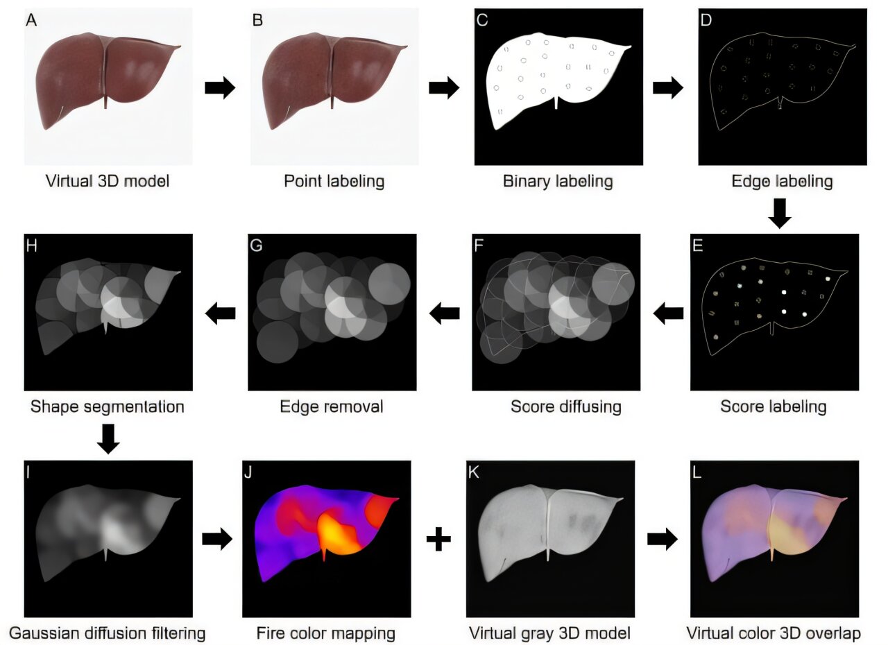
The flow diagram of mapping the virtual 3D distribution characters over the entire liver from PS-OCT and biopsy scores. Credit: Science Translational Medicine (2026). DOI: 10.1126/scitranslmed.adv7124
Each year, thousands of patients in the United States wait for a liver transplant, while transplant teams must make rapid, difficult decisions about whether donor organs are suitable for use. Many of those livers, particularly from older or medically complex donors, require careful evaluation before transplantation, and current assessments often sample only a small region of the organ.
In response, a team of University of Oklahoma researchers has developed a noninvasive imaging approach that could help transplant teams evaluate donor livers more comprehensively and make faster, more informed decisions. Qinggong Tang, Ph.D. leads the OU research lab that recently published these findings in Science Translational Medicine.
Doctors currently determine liver health primarily through biopsies, in which a small piece of tissue is removed and examined under a microscope. However, this is an invasive procedure and cannot account for the health of the entire liver. Tang said this can lead to cases in which doctors may not feel comfortable using a liver for a transplant if the biopsy results don't feel conclusive enough.