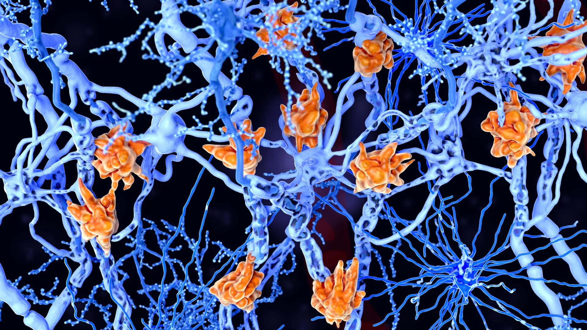
A cross-section of the spinal cord showing neurons and glial cells. (Juan Carlos Juarez Jaramillo/iStock/Getty Images Plus)
A diagnosis of multiple sclerosis (MS) comes with a wave of uncertainty about how the condition will progress.Now, new research points to a potential mechanism and treatment target for those who are most severely affected.MS damages nerve cells, stripping away their protective covering that keeps nerve signals firing.The new study, from researchers in the Netherlands, suggests that in the most severe cases of MS, an immune cell usually in charge of repairing damaged tissue and clearing away waste becomes overloaded with fat droplets.Known as "foamy microglia", these cells have been spotted in MS patients before, but it wasn't clear exactly what they were doing.According to the findings from this latest study, they could be key drivers of MS at its worst.Images of MS lesions with foamy microglia. Red arrows indicated fat-filled pockets of increasing size (left to right) inside the immune cells. (van der Vliet et al., Nat. Neurosci., 2026)MS is an autoimmune disease in which the body's immune system becomes overactive, mistakes its own cells as foreign, and starts causing damage through inflammation. But these foamy microglia suggest there's also more to the story."We found that patients with large numbers of these foamy microglia had a more severe disease course more frequently," says molecular physiologist Daan van der Vliet, from Leiden University in the Netherlands."It does not appear to be simply about the inflammatory response alone."The team analyzed post-mortem brain tissue from 28 people with secondary progressive MS, where the disease has progressed to the point where cognitive and physical function are declining.This tissue was compared against samples from 10 donated brains from people without the disease.The researchers compared brain tissue samples collected from people with and without severe MS, looking at the damage (lesions) on nerve cells. (van der Vliet et al., Nat. Neurosci., 2026)Using a combination of profiling techniques, the researcher created a map of proteins, fats, and active genes for the brain regions affected by MS lesions.These lesions form when the fatty, protective coating around nerve fibers, known as myelin, is attacked by immune cells that have become too aggressive.Not only was there a link between more foamy microglia and MS progression, but the researchers also found that the microglia were changing the mode of inflammation around the lesions – they had a different molecular signature in terms of proteins and enzymes.The researchers suggest that as microglia arrive to try and repair the damage done to neurons, they get clogged up with fats (beginning with myelin) and become overwhelmed, which in turn, makes the inflammation worse."These cells are probably trying to do something good: clearing up damage," says van der Vliet."But they become overloaded, so to speak. As a result, they can no longer effectively contribute to repair."Severe MS was associated with fat-laden, 'foamy' microglia. (Netherlands Institute for Neuroscience)The researchers also used a mouse model of MS, blocking one of the enzymes most active in foamy microglia. Tissue healing improved in these mice, further emphasizing the connection between these immune cells and worse MS progression.We're still in the early stages of this research, and clinical trials with MS patients will be needed to see if the foamy microglia link holds up.Researchers will also need to look at how these lesions that aren't repaired continue to develop over time.However, these are promising findings in terms of figuring out why some people with MS live relatively normal lives for decades, while others become paralyzed sooner or develop more severe symptoms at a young age.The study team is hopeful that the findings could help develop new MS treatments that target fat metabolism in cells.There's also the potential, along with other lines of research, to identify more severe cases of MS at an earlier stage.The researchers found signs of fats associated with foamy microglia floating around in cerebrospinal fluid, which they say could be measured as a marker of the disease.Related: Scientists Identify Specific Bacteria Linked to Multiple Sclerosis"That opens the possibility of developing biomarkers in the future that could help doctors identify earlier which patients are at risk of rapid decline – and which treatment would suit them best," says van der Vliet.The research has been published in Nature Neuroscience.