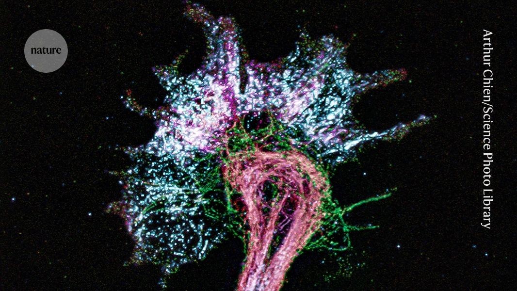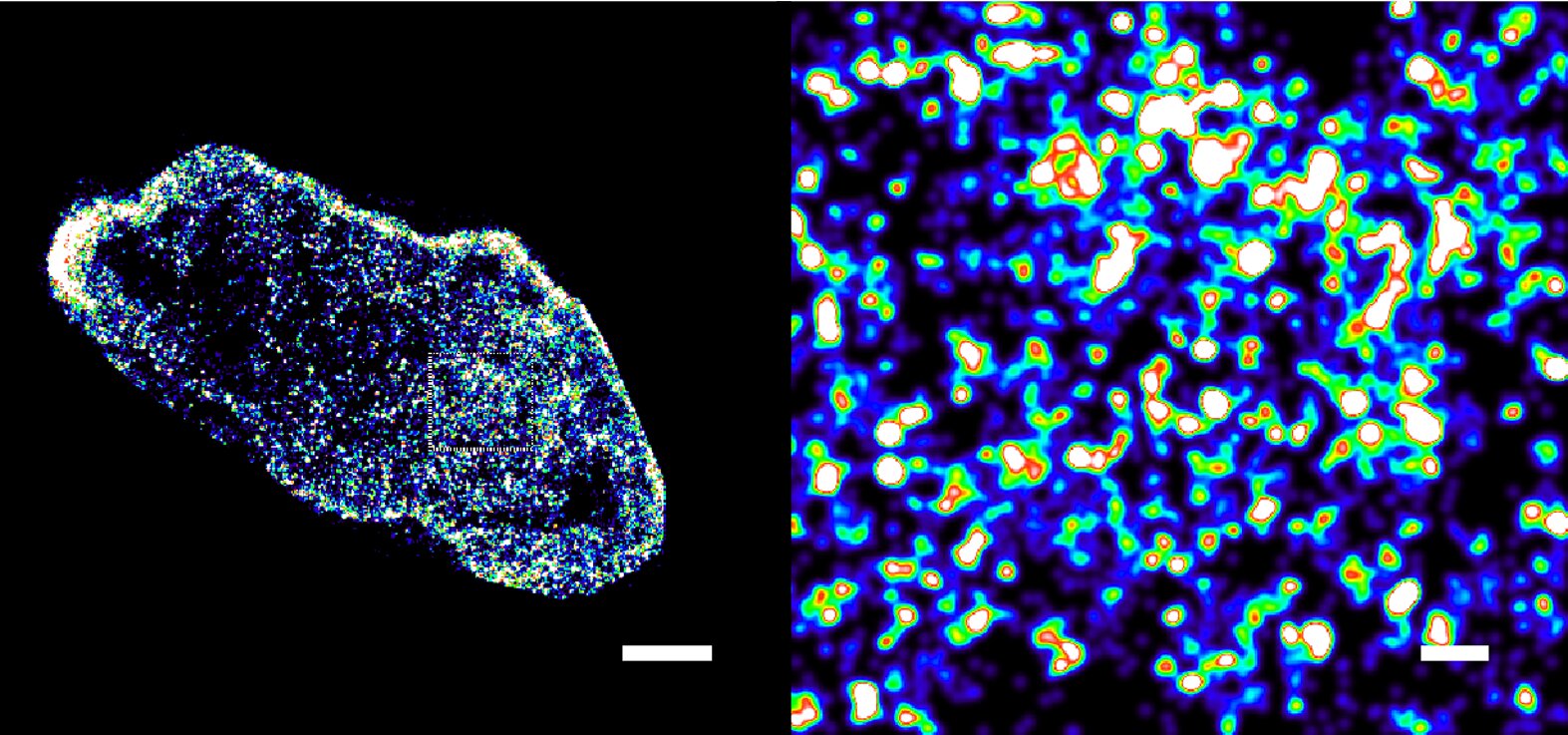
DNA inside a cancer cell from human bowl tissue, imaged at a scale of billionths of a metre. In the healthy cell, DNA is gathered into tight, distinct packages. In the cancer cell, it has come loose and spread out. Credit: Aiping Wang/Centro de Regulación Genómica/Guangdong Provincial People's Hospital
Researchers have developed fluorescent molecules that permit imaging of how DNA is packaged inside living cells at unprecedented resolution and, in preserved cells, at a resolution close to the width of the double helix itself.
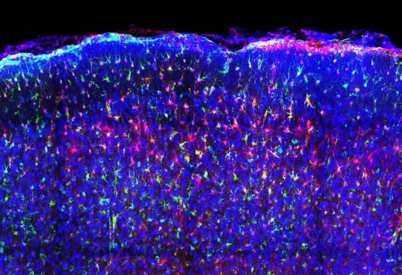
The team tested the fluorescent probes on slices of bowel tissue taken from three cancer patients. These are ordinary wax-preserved samples, which is how almost all hospitals store patient biopsies.
In the tumors, the DNA was noticeably looser and more spread out than in the healthy tissue sitting right beside it.
Other studies have found that DNA unpacks steadily as cancer takes hold, and the researchers suggest that how loosely a cell's DNA is folded could eventually serve as a measure of how far a tumor has progressed or how aggressive it is.