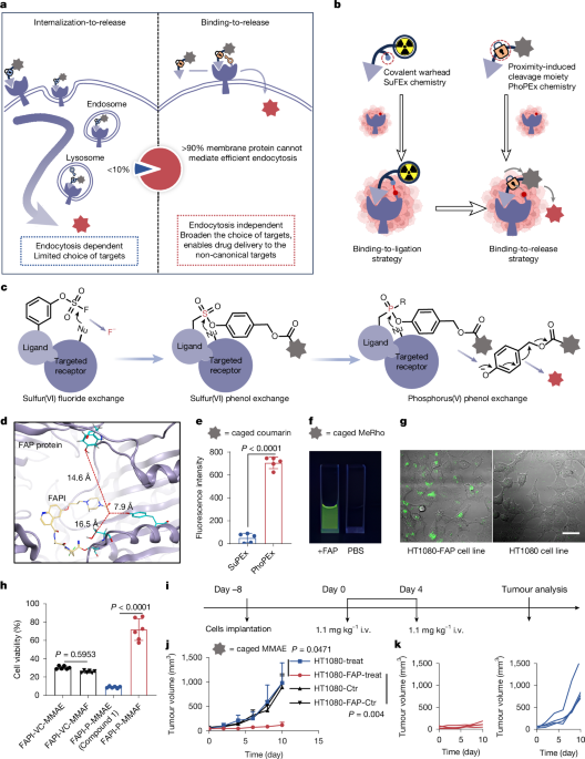
MainTargeted drug conjugates, such as antibody–drug conjugates (ADCs), represent a transformative therapeutic modality in oncology1,2,3. Although their clinical success is undeniable—with the market projected to reach US$26 billion8,9,10,11—the field remains constrained by a fundamental limitation: a strong dependency on efficient receptor-mediated endocytosis and lysosomal trafficking for payload release12,13 (Fig. 1a, left). This endocytic prerequisite creates a dual bottleneck. First, it restricts the druggable target landscape to a small fraction of the human membrane proteome (about 180 of more than 2,000 proteins4,5,6,7) that undergo efficient internalization, funnelling development into intense competition on a handful of antigens such as HER2 and TROP211,14. Second, it subjects drug delivery to the cumulative inefficiency of the multi-step lysosomal trafficking pathway, resulting in only a fraction of the internalized conjugates successfully releasing their payload15,16. Although recent evidence suggests that extracellular payload release may also contribute to ADC efficacy in some settings, the dominant pattern of the field and optimization efforts remain centred on endocytosis13,17. Consequently, the vast majority of tumour-specific cell surface antigens with poor internalization efficiency remain an untapped ‘blue ocean’, representing a substantial opportunity for expanding the therapeutic reach of drug conjugates.Fig. 1: PhoPEx chemistry enables a BTR strategy for endocytosis-independent targeted drug release.a, Schematic presentation of BTR compared with ITR strategy for drug delivery. b, Comparison of binding-to-ligation strategy and BTR strategy. c, Stepwise development of the PhoPEx reaction. d, Molecular docking of FAPI against FAP protein (PDB: 1z68), indicating the potential residue for PhoPEx reaction. e, Fluorescence intensity assay of FAPI-PhoPEx-coumarin and FAPI-SuPEx-coumarin were co-incubated with purified FAP protein (n = 5 independent samples). f, Representative photograph of the release of fluorescent molecules on FAP protein addition in vitro. FAPI-PhoPEx-MeRho (500 nM) and purified FAP (2 μM) were incubated in PBS (pH 7.4) at 37 °C, whereas the control group contained only 500 nM FAPI-PhoPEx-MeRho without FAP protein. g, Confocal fluorescence microscopy imaging of FAP-mediated release. HT1080-FAP cells and HT1080 control cells were co-incubated with FAPI-PhoPEx-MeRho for 24 h. Scale bar, 50 μm. h, Cell viability assay of HT1080-FAP cells that were treated with conjugates (50 nM) incorporating either the PhoPEx linker or a classical Val-Cit-linker. Cell viability was assessed following treatment with each linker conjugated to either MMAE or MMAF (n = 6 biological replicates). i, Schematic of the therapeutic dosing regimen (compound 1). j, Tumour growth curves in mouse models bearing HT1080-FAP or HT1080 tumours (n = 4 mice per group). k, Individual tumour growth curves. Data are shown as mean ± s.d. (e,h) and mean + s.d. (j). Statistical analysis was performed using a two-tailed unpaired Student’s t-test (e), one-way ANOVA followed by Tukey’s post hoc test (h) and two-way ANOVA followed by Tukey’s post hoc test (j). Data are representative of two (f,g) independent experiments. Illustrations in b created in BioRender. Liu, Z. https://BioRender.com/mvilhoy (2026).Source dataThe case of FAP exemplifies this untapped potential. As a pan-cancer antigen highly expressed on cancer-associated fibroblasts with minimal normal tissue expression18, its remarkable targeting potential is clinically validated by the diagnostic success of FAPI-based radiopharmaceuticals across more than 28 cancer types19. However, the development of FAP-targeted drug conjugates, such as the clinical candidate OMTX70520, has been hampered by the low internalization efficiency of FAP21, as evidenced by a best response of only disease stabilization in 26% of patients22. This impasse underscores the unmet need for a drug release pattern that bypasses the endocytic machinery.Inspired by the principles of ligand-directed chemistry23,24, which uses target binding to enable proximity-induced ligation25,26 (Fig. 1b), we developed a binding-to-release (BTR) strategy for endocytosis-independent drug delivery. Our core design involves engineering a conjugate with a new linker that remains stable in circulation. However, on ligand–receptor engagement, the linker can release the payload when positioned in proximity to nucleophilic residues within the binding pocket of the target. This proximity-induced reaction triggers the efficient, endocytosis-independent release of a membrane-permeable cytotoxic payload directly at the tumour site (Fig. 1a, right), thereby circumventing the endocytic bottleneck.PhoPEx enables binding-to-releaseTo translate this concept into reality, a chemical tool that combines circulatory stability with rapid, proximity-triggered cleavage is essential. Although sulfur(VI) fluoride exchange (SuFEx) chemistry offers a promising balance of stability and reactivity27,28, we initially sought to integrate a self-immolative phenol-based scaffold—a structure widely used to mediate efficient drug release29—by developing a sulfur–phenol exchange (SuPEx) system. However, unlike SuFEx, SuPEx—even with p-nitrophenol as the leaving group—showed negligible tyrosine reactivity in tubes (Extended Data Fig. 1a). Inspired by the review in ref. 30, we substituted sulfur (S) with phosphorus (P) as the central electrophilic atom and developed the phosphorus(V)–phenol exchange (PhoPEx) reaction (Fig. 1c), and this replacement enhanced the tyrosine reaction rate in vitro. Therefore, we engineered the first-generation PhoPEx linker, using p-hydroxybenzyl alcohol as the leaving group and conjugated it to a FAP-targeting ligand (FAPI) to evaluate its BTR ability. This design was based on molecular docking predictions indicating the proximity of reactive nucleophilic residues to the FAPI binding pocket within FAP (Fig. 1d). In vitro, PhoPEx-based BTR conjugates bearing caged fluorescent reporters (coumarin or MeRho) demonstrated FAP-dependent payload release after 24 h of co-incubation, a response absent in SuPEx-based conjugates and phosphate-buffered saline (PBS) controls (Fig. 1e,f). This specificity was confirmed in cellular models: HT1080-FAP exhibited pronounced fluorescence activation after 24 h, whereas HT1080 controls showed minimal signal (Fig. 1g).To investigate whether the BTR mechanism bypasses endocytosis, we compared the efficacy of conjugates bearing membrane-permeable (monomethyl auristatin E, MMAE) and comparatively impermeable (monomethyl auristatin F, MMAF) payloads. Although the internalization-to-release (ITR) Val-Cit-linker mediated cell killing in HT1080-FAP cells with both MMAE and MMAF, the PhoPEx-based BTR conjugate induced potent cytotoxicity only with MMAE; the MMAF-conjugate was ineffective (Fig. 1h). This disparity indicates that the BTR platform operates by an extracellular, non-internalizing release mechanism, as only the membrane-permeable MMAE could diffuse into cells to exert its effect. We acknowledge that this mechanism inherently necessitates the use of payloads with high membrane permeability. But this requirement aligns with the prevailing trend in ADC development2, driven by the success of agents such as DS8201 (Enhertu), in which highly permeable payloads such as DXd and MMAE are prioritized for their enhanced efficacy, particularly against heterogeneous tumours31. Furthermore, for a stromal target such as FAP, which is often expressed on cancer-associated fibroblasts rather than directly on cancer cells, an extracellular release mechanism offers a distinct advantage. The released, permeable drug can diffuse throughout the tumour microenvironment, potentially achieving a broader distribution and mitigating the challenge of tumour heterogeneity compared with an internalization-dependent approach (Supplementary Fig. 15). Tail vein administration of the MMAE-loaded BTR conjugate (compound 1, Extended Data Fig. 5b) elicited potent and specific tumour growth inhibition in HT1080-FAP xenografts, with negligible efficacy in FAP-negative HT1080 tumours (Fig. 1i–k). These initial results demonstrated that the first-generation PhoPEx linker enabled FAP-specific payload release without endocytosis. However, its suboptimal reaction efficiency still remained incompatible with the short circulatory half-life of small-molecule agents, necessitating the development of more reactive PhoPEx linkers.Given the suboptimal reaction kinetics of the first-generation linker, we systematically pursued structural optimization of the PhoPEx scaffold to enhance its reactivity while preserving stability. Based on the electronic (electron-withdrawing and electron-donating) effects of substituents, we introduced various functional groups at different positions on the benzene ring of the leaving group, designing and synthesizing a total of 15 PhoPEx structures (Extended Data Fig. 1c). Reactivity was assessed by their reaction with the tyrosine analogue (Tyr-OMe-NAc) in DMF with tetramethylguanidine (TMG) as a catalyst, whereas stability was evaluated by measuring hydrolysis rates in PBS buffer at pH 9.5 (Fig. 2a). The SuFEx structure was used as a benchmark for parallel comparison of both reactivity and stability. Using second-order kinetic fitting (Extended Data Fig. 1b), we obtained the reaction rate constants (kreact) and hydrolysis rate constants (khydro) for all 15 PhoPEx structures as well as the SuFEx reference (Fig. 2b and Extended Data Fig. 1e–j). When plotted with SuFEx as the baseline, PhoPEx structures falling within the red region exhibited reactivity comparable to SuFEx while demonstrating higher stability (Fig. 2c). Among these, the para-amide and meta-fluoro substitution performed best, with reaction rate constants of 4.35 × 10⁻³ M−1 s−1 and 3.92 × 10⁻³ M−1 s−1, respectively (SuFEx: 3.99 × 10⁻³ M−1 s−1), and hydrolysis rate constants of 3.07 × 10⁻⁸ M−1 s−1 and 2.28 × 10⁻⁸ M−1 s−1, respectively (SuFEx: 9.51 × 10⁻⁸ M−1 s−1) (Extended Data Fig. 1k).Fig. 2: Binding-induced PhoPEx release through Tyr745 proximity.a, Screening workflow for evaluating the reactivity and stability of PhoPEx-based compounds. b, High-performance liquid chromatography analysis of the reaction of PhoPEx-pCOOMe with tyrosine analogues, in which the reactant disappeared and both ligation and release products appeared. c, Screening results of 15 distinct PhoPEx variants, plotted against the SuFEx. d, In vivo PET-CT imaging of PhoPEx-FAPI-H and PhoPEx-FAPI-mF in HT1080-FAP tumour-bearing mice. e, Quantitative analysis of PET-CT results (n = 4 mice). f, Release efficiency of optimized compound 2 compared with precursor compound 1 after incubation with purified FAP protein; n = 3 independent samples. g, Workflow for determining the binding mode of the PhoPEx probe to FAP, integrating tandem mass spectrometry (MS/MS), site-directed mutagenesis and cryo-electron microscopy. h, MS/MS identification of Tyr745 on FAP as the primary reaction site. i, In vitro irreversible covalent binding assay of PhoPEx-FAPI-pCONH to FAP and mutant FAP (Y745F). RLC, radionuclide–ligand conjugate. j, Evaluation of drug release efficiency (compound 5) on engagement with wild-type compared with Y745F-mutant FAP for 30 min; n = 3 independent samples. k, Cryo-EM experiment for the density map showing the binding interface of the PhoPEx probe with FAP. The targeting moiety FAPI was covalently linked to the enzymatic Ser624. l, Molecular dynamics simulation and cryo-EM model of the PhoPEx probe bound to FAP. Data are shown as mean ± s.d. (e,f,j); statistical analysis was performed using a two-sided linear mixed-effects model, and no post hoc multiple comparisons were performed (f), and a two-tailed unpaired Student’s t-test (j). Data are representative of two (i) independent experiments and four mice (d). Illustrations in a and g created in BioRender; Liu, Z. https://biorender.com/mvilhoy (2026).Source dataIncorporation of the evolved meta-fluoro-substituted PhoPEx linker (compound 2, Extended Data Fig. 5b) markedly improved reactivity over the parent compound 1, resulting in a shortened reaction half-life of 3.5 h for better pharmacokinetic matching with small molecules, while largely preserving hydrolysis stability (Fig. 2f). In parallel, we prepared targeted covalent radiopharmaceuticals incorporating the evolved PhoPEx linkers (PhoPEx-FAPI-mF and PhoPEx-FAPI-pCONH) as well as the unsubstituted one (PhoPEx-FAPI-H), allowing for a comparison in vivo. Positron emission tomography–computed tomography (PET-CT) imaging showed that the PhoPEx-FAPI-mF achieved about two-fold higher tumour uptake in both SUVmax and SUVmean compared with the unsubstituted counterpart, indicating enhanced reaction efficiency within living systems (Fig. 2d,e). For PhoPEx-FAPI-pCONH, it demonstrated rapid and irreversible covalent binding to the FAP protein (Fig. 2i, left) and exhibited comparable tumour uptake against SuFEx-FAPI (Supplementary Figs. 8 and 24), suggesting that PhoPEx chemistry is comparable to SuFEx chemistry. The covalency led to higher tumour uptake and retention compared with the non-covalent FAPI04 (Extended Data Fig. 2f–i and Supplementary Fig. 11).BTR is mediated by residue proximityTo explore the underlying reaction mechanism on FAP protein—and specifically to rule out the possibility that payload release was mediated by the inherent hydrolytic activity of FAP, which would diverge from our proposed proximity-induced cleavage—we used tandem mass spectrometry (MS/MS), site-directed mutagenesis and cryo-electron microscopy (cryo-EM) to identify the key nucleophilic residue responsible for the reaction (Fig. 2g). Tandem MS analysis indicated that Tyr745 was the primary reaction site after incubation of [NatLu]Lu-PhoPEx-FAPI-pCONH with FAP protein (Fig. 2h), with minor secondary sites located on adjacent residues within the same α-helix, such as His743 and Thr741. Mutation of Tyr745 to phenylalanine (Phe) (Supplementary Fig. 4) reduced covalent binding efficiency (Fig. 2i, right), resulting in behaviour similar to the reversible covalent inhibitor FAPI04 (Extended Data Fig. 2e), and the payload release rate decreased by 63% compared with that of wild-type FAP (Fig. 2j). Finally, we used cryo-EM to further resolve the binding mode. Owing to the rapid reaction kinetics of the meta-fluoro derivative, which hindered capture of the initial binding state, we substituted it with an ortho-methyl derivative as an analogue to slow the process. The cryo-EM density map showed electron density (blue mesh) for the FAPI targeting head bound to FAP, whereas the DOTA and MMAE moieties exhibited high flexibility and thus lacked well-defined density. Combined with molecular dynamics simulations, the results indicate that the reactive phosphorus centre is spatially adjacent to Tyr745, consistent with previous experimental findings (Fig. 2k,l and Supplementary Fig. 20).BTR is target-specific and efficientHaving established the PhoPEx-based BTR strategy and its proximity-induced cleavage mechanism in model systems, we next asked whether this approach functions in clinical samples. To address this, we developed an activatable fluorescent probe, FAPI-P-Cy5-QSY21. On binding to FAP, the probe releases the quencher QSY21, restoring Cy5 fluorescence32,33 (Extended Data Fig. 3a). FAPI-P-Cy5-QSY21 showed FAP-specific fluorescence enhancement in vitro, in cells and in mouse models (Extended Data Fig. 3b–e). We then evaluated its performance on clinical specimens. Intraoperative assessment of lymph node metastasis in medullary thyroid carcinoma at present relies on subjective selection of a few nodes for frozen section analysis, which limits diagnostic accuracy. Given that FAP co-localizes with tumours in this cancer34, we proposed that incubating all resected lymph nodes with the probe and measuring fluorescence intensity could objectively determine FAP expression in metastases (Extended Data Fig. 3f). After optimizing probe concentration, incubation time and temperature (Extended Data Fig. 4d–f and Supplementary Fig. 5), we bisected lymph nodes (typically <5 mm in diameter), disrupted their capsules, incubated them with 2 μM probe at 37 °C for 40 min and washed three times with PBS. This protocol produced two distinct fluorescence intensity populations (Supplementary Fig. 6). Subsequent FAP immunohistochemistry (IHC) showed that nodes with higher fluorescence intensity were mostly FAP-positive, whereas those with lower intensity were predominantly FAP-negative (Extended Data Fig. 3g). Quantitative analysis showed that the average radiant efficiency in FAP-positive nodes was 2.9-fold higher than in FAP-negative nodes (Extended Data Fig. 3h). Together, these results demonstrate that the PhoPEx-based BTR strategy is effective in clinical specimens.With the BTR platform validated, we then used meta-fluoro-substituted PhoPEx linker to continue evaluating whether BTR would meet the critical parameters for a controlled-release system—specificity and efficiency—and exploring its potential advantages compared with the classical internalization-to-release (ITR) pattern (exemplified by the Val-Cit dipeptide linker35 used in the approved ADCs) when both are applied to the FAP target. Specificity and efficiency are two parameters that are central to controlled-release systems: high specificity minimizes off-target effects, reduces systemic toxicity and increases the maximum tolerated dose (MTD); high efficiency ensures sufficient drug release within tumour tissues, enhancing efficacy and lowering the minimum effective dose. At the molecular level, FAP-BTR-SMDC (compound 5) exhibited high binding selectivity for FAP protein, showing more than 1,000-fold selectivity against homologous proteins (Fig. 3b). Even in the presence of 100-fold equivalents of nucleophilic amino acids, 10-fold equivalents of other proteins (for example, albumin, alkaline phosphatase), or in human serum (Extended Data Fig. 5d,e), payload release—ranging from fluorescent molecules (compound 3) to cytotoxic agents such as the microtubule inhibitor MMAE (compound 5) and the TOPI inhibitor exatecan (compound 4)—occurred rapidly and specifically only on addition of FAP protein (Fig. 3c,d, Extended Data Fig. 5b and Supplementary Figs. 16, 25 and 26). Notably, Gly-Pro dipeptide, a common FAP-release substrate, can be non-specifically cleaved by homologues, such as DPPIV and PREP, as well as hydrolases, such as CTSB36 (Extended Data Fig. 5f,g). At the cellular level, FAP-BTR-SMDC (compound 5) demonstrated enhanced cytotoxicity against FAP-positive cells, with IC50 values of 498.9 nM in U87MG-Vector and 9.0 nM in U87MG-FAP, suggesting release specificity (Fig. 3e and Supplementary Fig. 14). Compared with the ITR Val-Cit-linker, compound 3 (BTR PhoPEx linker) released more than eight-fold higher fluorescent signal at the same time point under equivalent incubation conditions, indicating better release efficiency (Fig. 3f,g). In animal models, tumour analysis following intravenous administration of 1.0 mg kg−1 FAP-BTR-SMDC (compound 5) showed that MMAE concentrations in HT1080-FAP tumours were 34.5-fold higher than in HT1080 tumours at 24 h (Fig. 3i), and MMAE exposure (area under the curve from 0 to 120 h, AUC0–120 h) in HT1080-FAP tumours was 5.9-fold higher for FAP-BTR-SMDC (compound 5) than for FAP-ITR-SMDC (Val-Cit-linker, equal moles of MMAE), and comparable to FAP-ITR-ADC (FAP-targeted sibrotuzumab coupling with VC-MMAE through cysteine and maleimide, drug-to-antibody ratio (DAR) of 3.5, Extended Data Fig. 6b–d, equivalent dose of MMAE) (Fig. 3m). Single photon emission computed tomography with computed tomography (SPECT-CT) imaging showed additional uptake of FAP-BTR-SMDC in the liver. Thus, liver and blood were used as reference normal tissues to assess nonspecific release (Fig. 3h and Supplementary Fig. 10). Both in blood and in liver, nonspecific release in non-target organs was lower for FAP-BTR-SMDC, with tumour-to-blood and tumour-to-liver ratios reaching 39.7 and 358.1, respectively—14.7- and 55.1-fold higher than those of FAP-ITR-SMDC, and 3.6- and 58.7-fold higher than those of FAP-ITR-ADC (Fig. 3j–l,n and Supplementary Fig. 27). These results indicate that the PhoPEx linker operating through the BTR mechanism showed improved release specificity and efficiency compared with the internalization-dependent Val-Cit-linker on FAP protein. Moreover, despite the different pharmacokinetic profiles, the FAP-BTR-SMDC achieved tumour drug exposure comparable to that of the antibody-based FAP-ITR-ADC, with higher tumour-to-normal tissue ratios.Fig. 3: The BTR strategy exhibits release specificity for efficient drug delivery.a, Workflow for evaluating release specificity and efficiency. b, Assessment of the binding selectivity of compound 5 for FAP protein relative to homologous proteins (n = 6 technical replicates for FAP and 4 technical replicates for DPPIV and PREP). c, Release specificity test of compound 3 by incubation with nucleophilic amino acids (100 equiv.) and different proteins (10 equiv.) DPP IV, dipeptidyl peptidase IV; PREP, prolyl endopeptidase; ALB, albumin; ALP, alkaline phosphatase; CTSB, cathepsin B; n = 3 independent samples. d, Evaluation of payload release capacity of BTR strategy, including MMAE, exatecan and MeRho (compound 3); n = 3 independent samples. e, Cytotoxicity of compound 5 in U87MG-FAP compared with U87MG-Vector cell lines; n = 6 biological replicates. f, Confocal microscopy imaging comparing payload release between compound 3 and FAPI-VC-MeRho in HT1080-FAP cells. Scale bar, 50 μm. g, Fluorescence quantification from confocal data (n = 3 independent experiments). h, SPECT-CT imaging of HT1080-FAP tumour-bearing mice after [177Lu]Lu-compound 5 administration (n = 3 mice). i, MMAE concentration analysis in HT1080-FAP and HT1080 tumours 24 h after intravenous injection of compound 5 (1.0 mg kg−1; n = 4 mice per group). j–l, Pharmacokinetics of released MMAE in tumour, blood and liver for FAP-BTR-SMDC (j), FAP-ITR-SMDC (k) and FAP-ITR-ADC (l) in the HT1080-FAP model; n = 4 mice per time point. m,n, Area under the curve (AUC) values calculated from MMAE concentrations (m) and tumour-to-normal (T/N) ratios (n); n = 4 groups of mice (each group contained 6 mice euthanized at 2, 6, 24, 48, 72, 120 h for biodistribution). T/B, tumour-to-blood; T/L, tumour-to-liver. Data are mean ± s.e.m. (b), mean ± s.d. (c–e,g,i,m,n) and mean + s.d. (j–l). Statistical analysis: two-tailed unpaired Student’s t-test (i,g), and one-way ANOVA followed by Tukey’s post hoc test (c). Data are representative of three independent experiments (f) and three mice (h). Illustrations in a created in BioRender; Liu, Z. https://biorender.com/mvilhoy (2026).Source dataBTR broadens therapeutic windowWe then tested whether release specificity and efficiency would translate into a broadened therapeutic window. In acute toxicity studies, in which the MTD was defined as the highest dose that did not cause body weight loss exceeding 20%, the MTD of the FAP-BTR-SMDCs was 7.5-fold molar dose higher than that of the FAP-ITR-SMDCs, indicating a reduction in systemic toxicity (Fig. 4a). In cell-derived xenograft (CDX) model efficacy studies, at equivalent doses, FAP-BTR-SMDCs demonstrated greater tumour growth inhibition. A single intravenous administration of FAP-BTR-SMDCs (6.2 mg kg−1) resulted in complete response in 100% of mice (8 out of 8) within 30 days, accompanied by stable body weight and 100% survival (Fig. 4b–f and Supplementary Figs. 13 and 29). In patient-derived xenograft (PDX) models, we evaluated FAP-BTR-SMDC against FAP-ITR-SMDC and a reference FAP-ITR-ADC. In a sarcoma PDX (Fig. 4h–j), in which FAP is expressed directly on tumour cells, FAP-BTR-SMDC (dosed at 6.2 mg kg−1, about 15% of its MTD) showed better anti-tumour efficacy than FAP-ITR-SMDC and achieved efficacy slightly better than the FAP-ITR-ADC (dosed at 22.2 mg kg−1, approximately 50% of its MTD; Extended Data Fig. 6e). In a pancreatic adenosquamous carcinoma (PASC) PDX (Fig. 4k–m)—a clinically aggressive, treatment-refractory malignancy characterized by a dense desmoplastic stroma that limits the penetration of large molecules37—FAP is expressed on cancer-associated fibroblasts rather than on tumour cells. In this model, FAP-BTR-SMDC demonstrated better efficacy compared with the FAP-ITR-ADC. Terminal tumour weight measurements and photos (Extended Data Fig. 7e) of tumours indicated the improved efficacy of FAP-BTR-SMDC (tumour weight: FAP-BTR-SMDC group 0.19 ± 0.07 g compared with ADC group 1.32 ± 0.41 g). Apart from these models, FAP-BTR-SMDC also showed anti-tumour activity in MC38-FAP CDX (Extended Data Fig. 7b), hypopharyngeal PDX (Extended Data Fig. 7f–j) and periampullary carcinoma PDX models (Extended Data Fig. 7k–n), with efficacy correlating with FAP expression levels. Collectively, these data from both toxicity tolerance and treatment efficacy dimensions indicate that the BTR platform broadens the therapeutic window.Fig. 4: The BTR drug delivery system exhibits improved therapeutic profiles on FAP protein in CDX and PDX models.a, Evaluation of the MTD for FAP-BTR-SMDC (compound 5) and FAP-ITR-SMDC in healthy CD-1 mice (n = 10 mice per group). b, The regimen for the CDX treatment study. c, Tumour growth curves in the CDX model across treatment groups, FAP-BTR-SMDC (compound 5, 1.0/2.1/6.2 mg kg−1, equivalent to 10/20/60 nmol per mouse) and FAP-ITR-SMDC (0.85/1.7 mg kg−1, equivalent to 10/20 nmol per mouse). d, Body weight of mice monitored during the CDX model treatment period. e, Survival curves for the different treatment groups in the CDX model. f, Individual tumour growth curves for each mouse in the CDX model groups. g, The regimen for the PDX treatment study. h, PET-CT image and IHC staining for FAP expression in the sarcoma PDX model. Scale bar, 250 μm. i, Tumour growth curves in the sarcoma PDX model. j, Body weight change ratio of mice during the sarcoma PDX model treatment period. k, PET-CT image of the PDX model and IHC staining for FAP expression in the PASC PDX model. Scale bar, 250 μm. l, Tumour growth curves in the PASC PDX model. m, Body weight changes of mice during the PDX model treatment period; n = 8 mice per group (c,d,e,i,j) and n = 6 mice per group (l,m). Data are shown as mean + s.e.m. (c); mean ± s.d. (a) and mean + s.d. (d,i,j,l,m). Statistical analysis was performed using a two-sided mixed-effects model. P-value is for the group main effect; no post hoc multiple comparisons were performed (c) and two-way ANOVA followed by Tukey’s post hoc test (i,l). Data are representative of two mice (h,k). Illustrations in b and g created in BioRender; Liu, Z. https://biorender.com/mvilhoy (2026).Source dataThe rational design of BTR strategy on FAP has demonstrated considerable potential, while also revealing vast, untapped chemical opportunities at the protein-binding interface. mRNA display is a high-throughput screening technology that can create ultralarge libraries (typically exceeding 1013 unique members) and can support highly efficient direct in vitro screening38 (Fig. 5a). In previous work, researchers reported the identification of covalent macrocyclic peptides containing SuFEx through mRNA display screening39. Given the mechanistic similarities between PhoPEx and SuFEx chemistry, we directly substituted the SuFEx-based unnatural amino acid in the cyclic peptide with an unnatural amino acid incorporating the PhoPEx release structure and conducted systematic evaluation (Fig. 5b). Experimental results showed that the payload release rate reached its maximum when the linker contained four carbon atoms, with a rate constant of 3.25 h−1, enabling complete release of the fluorescent molecule within 1 h (Fig. 5c,d). This preliminarily suggested the feasibility of screening PhoPEx-based peptide–drug conjugates (PDCs) by mRNA display.Fig. 5: Expand the BTR system to other clinically important targets.a, Schematic of the rational design approach and an mRNA display-based pipeline for discovering covalent macrocyclic peptides that facilitate the BTR strategy. b, Structural model of a representative SuFEx-based covalent macrocyclic peptide and its BTR version incorporating PhoPEx chemistry. c, Release kinetics of fluorescent molecules as a function of linker chain length. d, Time-lapse fluorescence imaging of cyclopeptide–PhoPEx–coumarin after addition of FAP protein. e, Structural analysis of ligand and PD-L1 protein. f, Rational design of BTR strategy on PD-L1. g, Fluorescence intensity assay of different compounds (substituted from Gly or Leu), which were incubated with purified PD-L1 protein; n = 3 independent samples. h, Representative photograph of the release of fluorescent molecules on PD-L1 protein addition in vitro. PD-L1-PhoPEx-MeRho (PD4) and purified PD-L1 protein were co-incubated in PBS (pH 7.4), whereas the control (Ctrl) group contained only PD-L1-PhoPEx-MeRho without PD-L1 protein. i, Release kinetics of MMAE on PD-L1 added; n = 3 independent samples. j, MMAE concentration analysis in HT1080 tumours 24 h after intravenous injection of PD-L1-BTR-SMDC (with or without injection of BMS-986189 1 mg per animal) and PD-L1-ITR-SMDC (all 10 nmol per mouse; n = 4 mice per group). k–m, PET-CT image and IHC characterization of PDX models (k), tumour growth curve (l) (4.2 mg kg−1 for PD-L1-BTR-SMDC, 4.8 mg kg−1 for PD-L1-ITR-SMDC, both equivalent 20 nmol per mouse; n = 6 mice per group) and tumour weight on day 16 (m). Scale bar, 250 μm. Data are shown as mean ± s.d. (g,i,j,m) and mean + s.d. (l). Statistical analysis was performed using one-way ANOVA followed by Tukey’s post hoc test (j,m) and two-way ANOVA followed by Tukey’s post hoc test (l). Data are representative of two independent experiments (d,h) and two mice (k).Source dataBTR applies to PD-L1-targeted drug deliveryFurthermore, we performed a bioinformatic analysis of all ligand–protein co-crystal structures in the PDBbind database40,41,42 (v2020.R1, 19,037 entries), which showed that 17,967 structures (94.4%) contain at least one tyrosine residue within 10 Å of the ligand. PD-L1 was selected as the second target because of its clinical importance and poor internalization. According to a reported cyclic peptide ligand43 (PDB: 9MAP), we introduced PhoPEx at either a Gly or a Leu position (Fig. 5e,f). Substitution at Gly reduced binding affinity to 0.54 ± 0.11 μM, whereas substitution at Leu maintained high affinity (8.4 ± 2.2 nM) (Extended Data Fig. 8b and Supplementary Fig. 28). The high-affinity variant (Leu) enabled better PhoPEx-mediated release of coumarin, MeRho and MMAE in vitro (Fig. 5g–i). In vivo, in HT1080 tumour-bearing mice, which expressed PD-L1 (Extended Data Fig. 8d), the PD-L1-BTR-SMDC conjugate showed more tumour-specific MMAE release than PD-L1-ITR-SMDC and could be blocked by the peptide BMS-986189 (Fig. 5j). The PD-L1-BTR-SMDC, therefore, showed better anti-tumour efficacy in a PDX model of gastric adenocarcinoma (Fig. 5k–m and Extended Data Fig. 8c). These findings suggest that the BTR strategy holds promise for extension to other important therapeutic targets with versatile routes, demonstrating broad potential applicability.In this study, we first developed the PhoPEx chemistry, optimized its reactivity and stability, and preliminarily studied its mechanism (Supplementary Fig. 17). Based on PhoPEx chemistry, we developed the BTR strategy, which enables payload release independently of endocytosis. Using FAP as a model target with poor internalization21 (Extended Data Fig. 5h,i), we optimized the PhoPEx linker and identified Tyr745 as the key nucleophilic residue for the proximity-induced reaction. The platform was successfully applied to SMDCs, activatable fluorescent probes, and—on a second target—a PD-L1-targeted conjugate, demonstrating its adaptability.The BTR strategy is intended as a complementary approach to the conventional ITR strategy. ITR is highly effective for targets with efficient endocytosis (for example, HER2, TROP2), but faces inherent limitations for most of the membrane proteins that internalize poorly. Recognizing this gap, several recent approaches have explored non-internalizing or extracellular release mechanisms13,17,44,45,46, including releasing payload through extracellular cathepsin cleavage, by tumour-microenvironment proteases, or by developing a two-component click-to-release system using an exogenous trigger to cleave the linker. Our BTR platform uses a single-agent, proximity-induced chemical reaction directly triggered by target binding, without relying on enzymatic activity or exogenous co-administration.Compared with intracellular payload release, extracellular release of a membrane-permeable payload could offer potential advantages, including better drug distribution within the tumour microenvironment and the ability to kill neighbouring antigen-negative cells (bystander effect). At the same time, it could in principle raise off-tumour toxicity concerns. Yet, this risk is manageable, as it is suggested that the amount of payload released in the tumour represents only a small fraction of the total administered dose47, and the side effects caused by the minor leakage from the tumour may be insignificant. Moreover, potential mitigation strategies include optimizing payload permeability, adopting fractionated dosing regimens and implementing rigorous haematological monitoring, which are also available in preclinical or clinical settings. Looking forward, the PhoPEx-based BTR platform operates through a direct binding-induced mechanism that transcends the cellular uptake limitations of conventional approaches. Thus, the BTR strategy provides a complementary framework for targeting antigens that are not efficiently addressed by ITR, expanding the scope of conjugate therapies.MethodsSynthesis and isolation of all related compoundsAll available compounds were purchased from commercial suppliers around the world. All solvents for synthesis were purchased from local suppliers, and solvents of high-performance liquid chromatography (HPLC)-grade were purchased from Fisher Scientific. The synthetic routes and chemicals used for all related compounds are shown in Supplementary Information section 2 (Core PhoPEx structure was prepared following a reported method48). Both UPLC-mass spectrometry and high-resolution mass spectrometry were used to assess the quality of all compounds and are provided in Supplementary Information section S6.Proteins and antibodiesFAP (C14G) were purchased from Novoprotein. DPPIV (HY-P70017), PREP (HY-P703584), ALB (HY-P1956A) and ALP (HY-P2818) were purchased from MCE. CTSB (C6286) were purchased from Sigma. PD-L1 (PD1-H5229 and PD1-H82E5) were purchased from ACRObiosystems. Sibrotuzumabs were purchased from WuXi Biologics. Rabbit monoclonal anti-FAP antibody was purchased from Abcam, ab207178, clone EPR20021, 1:50; mouse monoclonal anti-CK (pan) antibody from ZSGB-BIO, ZM-0069, clone AE1/AE3, 1:100; mouse monoclonal anti-CK5/CK6 antibody from ZSGB-BIO, ZM-0313, clone OT1F8, 1:100; mouse monoclonal anti-CK7 antibody from ZSGB-BIO, ZM-0071, clone UMAB161, 1:100; and rabbit monoclonal anti-PD-L1 antibody from Abcam, ab205921, clone 28–8, 1:250.Chemical reaction rate detectionThe purified substrate was dissolved in DMF to prepare a stock solution, then diluted to a concentration gradient (2 mM to 0.2 mM). Each solution was analysed by UPLC-MS with ultraviolet detection; peak areas were integrated at λmax, and the process was repeated in triplicate to generate a standard curve. For reactivity assays, each reaction mixture (300 μl) contained 1.5 mM substrate, 6 mM tetramethylguanidine and 22.5 mM Tyr-OMe-NAc (15 equiv.) at 37 °C. At timed intervals, aliquots (30 μl) were withdrawn, vortexed, centrifuged and quenched with 30 μl of 40 mM formic acid in acetonitrile. Quenched samples were analysed by UPLC-MS; concentrations were determined from the standard curve. Pseudo-first-order kinetics were fitted to obtain second-order rate constants. For the faster substrates (nos. 11 and 12), conditions were adjusted to 1 mM substrate, 1 mM tetramethylguanidine and 2 mM Tyr-OMe-NAc (2 equiv.), and second-order kinetics were directly simulated. Stability evaluation was performed in pH 9.5 PBS containing 1.5 mM substrate, using the same detection method as in the reactivity assay.Detection of release experiments in vitroFor the experimental group, the molecular stock solution was diluted with PBS and then mixed with the protein solution at an appropriate ratio (protein:small molecule = 4:1 or 5:1). The mixture was incubated at 37 °C, and aliquots were withdrawn at designated time points. The concentration of the molecule was determined using a pre-established standard curve. For the control group, the molecular stock solution was diluted with PBS to the same concentration and incubated under identical conditions without protein. Each condition was performed in three independently prepared samples. Fluorescent molecules were detected using a microplate reader; molecules lacking strong characteristic absorption (for example, MMAE) were quantified by UPLC-MS.Human lymph node staining and IHCHuman lymph node tissue excised by clinicians was cleaned to remove the surrounding adipose tissue and bisected with a surgical blade. One half was paraffin-embedded, sectioned into 4 µm slices and stained with haematoxylin and eosin for histology. For IHC, sections were deparaffinized in xylene, rehydrated through graded ethanol and subjected to antigen retrieval by microwave treatment in citrate buffer (pH 6.0). The other half was immersed in a fluorescent molecule solution, agitated at 37 °C for 40 min, then washed three times with PBS (10 min at 37 °C with agitation for each wash). Finally, the tissue was imaged using a near-infrared fluorescence imaging system. The use of patients’ lymph nodes was approved by the Institutional Review Board of the Cancer Hospital, Chinese Academy of Medical Sciences, under approval no. 25/328–5274.Procedures for radiolabelling and PET/SPECT-CT imagingThe radiolabelling and imaging were in accordance with previous literature49. For radiolabelling, the reaction mixture was prepared in 0.2 M sodium acetate buffer (pH 4.0–4.5) containing the appropriate labelling molecule (10 nmol) and the corresponding radionuclide (177Lu, 68Ga or 86Y). The mixture was heated at 90–95 °C for 10–15 min and then allowed to cool. To purify the mixture, the cooled reaction product was filtered through a pre-conditioned Sep-Pak Light C18 cartridge using deionized water, with the aim of removing unbound radionuclides. The radiolabelled compound was subsequently eluted with ethanol. This eluate was then diluted with saline for use in follow-up experiments. Where needed, ethanol was eliminated by nitrogen blowing using a Termovap sample concentrator before the dilution step. Evaluation of radiochemical yield was conducted using a radioactivity meter, and radiochemical purity was analysed by radio-HPLC. PET/SPECT-CT imaging for animals was performed on a Mediso nanoScan PET 122S system (Mediso) or InliView-3000B PET/SPECT/CT imaging system (Novel Medical) with mice injected with compounds through the tail vein. Mice were anaesthetized with isoflurane in oxygen 10 min before each imaging time point. Standard data acquisition and image reconstruction of the PET or SPECT data were performed. Data analysis was performed on Interview Fusion software (v.3.09.008.0000) and NMSoft-AIWS software (v.1.8).Tumour xenograft construction and treatment experimentsFor CDX models targeting FAP, HT-1080 or HT-1080-FAP cells (5 × 10⁶ in PBS) were implanted subcutaneously into the right flank of 6–8-week-old female NU/NU nude mice, whereas MC38-FAP cells (5 × 10⁵ in PBS) were implanted into 4–6-week-old female C57BL/6 mice. For CDX models targeting PD-L1, HT-1080 cells (5 × 10⁶ in PBS) were implanted into 6–8-week-old female BNDG mice.For all PDX models, BNDG mice aged 6–8 weeks were used, with the sex matched to that of the patient from whom the tumour specimen was obtained. Primary patient tumour tissue was directly implanted subcutaneously to generate the P1 generation. Once the P1 tumour had grown to 1,000–1,500 mm3, it was harvested and cut into fragments of approximately 3 mm in diameter, which were then implanted by puncture into the forelimb axilla of recipient mice to establish the P2 generation. Subsequent passages were performed in a similar manner. The final generation used for treatment was subjected to PET imaging and IHC to evaluate target expression. The use of patients’ tissues for PDX modelling was approved by the Institutional Review Board of the Cancer Hospital, Chinese Academy of Medical Sciences, under approval no. 25/328-5274, and by the Institutional Review Board of Beijing Cancer Hospital, under approval no. 2026KT67.All animal experiments were performed in accordance with protocols approved by the Institutional Animal Care and Use Committee of Peking University, under approval no. CCME-LiuZB-2, and by the Institutional Animal Care and Use Committee of Chinese Institute for Brain Research, Beijing, under approval no. CIBR-IACUC-071. The tumour volume was computed using the formula: volume = (length × width2)/2. In none of the experiments did the tumour burden surpass 10% of mouse weight and 25% weight loss compared with the original weight (according to the limits defined by the IACUC protocol).Cell cultureThe HT1080-FAP cells customized by WuXi AppTec were cultured in Eagle’s minimum essential medium containing 10% fetal bovine serum (FBS), 1% antibiotic–antimycotic and 4 μg ml−1 blasticidin S. HT1080 cells (WuXi AppTec) were cultured in minimum essential medium containing 10% FBS, 1% antibiotic–antimycotic. The U87MG-FAP and U87MG-Vector (both from Pyrotech Biotechnology) were cultured in minimum essential medium containing 10% FBS, 1% antibiotic–antimycotic and 2 μg ml−1 puromycin. The MC38-FAP cells (WuXi AppTec) were cultured in Dulbecco’s modified Eagle medium containing 10% FBS, 1% antibiotic–antimycotic and 3 μg ml−1 puromycin. All cells were regularly tested for mycoplasma contamination and cultured or incubated (as in cell-based assays) in a 5% CO2 incubator at 37 °C.For the fluorescent assay, cells were seeded in eight-well confocal dishes and incubated with different compounds (FAPI-PhoPEx-MeRho at 10 μM for 24 h, compound 3 and FAPI-VC-MeRho at 2 μM). Fluorescence was observed at room temperature using a Nikon A1R-si Laser scanning confocal microscope. The fluorescence intensity was measured using ImageJ.Half-maximal inhibitory concentrationInitially, two types of cells were seeded into a 96-well plate at a density of 5,000 cells per well and cultured in an incubator maintained at 37 °C for 12 h. A series of drug solutions with concentration gradients was then prepared using complete cell culture medium. After aspirating the original medium, the drug solutions were added for treatment. Following 6 h (compound 5, mF substitution) to 12 h (compound 1, no substitution) of incubation, the drug solutions were removed, and the cells were gently washed with PBS. Fresh drug-free complete medium was then added, and the cells were cultured for an additional 24 h. Finally, cell viability was assessed using the CCK-8 reagent.Biological distribution experimentThe biological distribution experiments of each compound were evaluated by four groups of mice, each group containing 6 mice, which were euthanized at 2, 6, 24, 48, 72, and 120 h after injection for tissue collection. After tail vein administration of the drug, blood, liver and tumour tissues were collected and weighed. RIPA lysis buffer and PMSF were added, and the tissues were homogenized at 4 °C for 30 min (for about 500–900 mg tissue: 1 ml RIPA buffer containing 100 μl of 100 mM PMSF per ml of buffer). After lysis, 2 ml RIPA buffer, 2 ml methanol and 1 ml acetonitrile were added. The sample was sonicated for 10 min and centrifuged at 4,000 rpm for 10 min. A 5 ml aliquot of supernatant was evaporated using a centrifugal concentrator. The residue was reconstituted in 800 μl of water–methanol–acetonitrile (1:1:1, v/v/v) and filtered through a 0.22-μm membrane. Finally, MMAE was quantified by UPLC-MS based on mass spectrometry signal integration (Supplementary Fig. 9). The AUC for each group (6 mice) was calculated based on the payload concentrations determined by the aforementioned assays at the six indicated time points.Cryo-EM experimentFor single-particle cryo-EM analysis, FAP protein at a concentration of 0.4 mg ml−1 was incubated with a 20-fold molar excess of enantiomerically pure FAPI-oMe-MMAE for 2 h at 4 °C. The resulting complex was then prepared for cryo-EM using graphene-oxide-coated grids. Data collection was performed, followed by single-particle data processing, which ultimately led to three-dimensional reconstruction and model building (Supplementary Tables 1 and 2 and Supplementary Fig. 22).Surface plasmon resonance experimentSurface plasmon resonance (SPR) experiments were performed on a Biacore 8K+ (Cytiva) using a Series S Sensor Chip SA (Cytiva, 29104992). Biotinylated PD-L1 was captured onto the chip by streptavidin–biotin interaction at a flow rate of 30 μl min−1. Analytes were then injected in a single-cycle kinetics at six consecutive increasing concentrations (5.12–500 nM, approximately 2.5-fold serial dilution). Injection parameters consisted of a constant flow rate of 30 μl min−1, 150 s association and 600 s dissociation per concentration. Before each analyte injection, a buffer blank was injected under identical conditions. The resultant binding curves were fitted to a 1:1 binding model using Biacore Evaluation Software for each dataset. Each analyte was tested in triplicate.Reporting summaryFurther information on research design is available in the Nature Portfolio Reporting Summary linked to this article.
A binding-to-release strategy for targeted anticancer drug delivery - Nature
A binding-to-release drug conjugate strategy enables targeted payload release without cellular internalization, improving tumour specificity, efficacy and expanding therapeutic targets beyond conventional drug conjugates.
10,017 words~46 min read