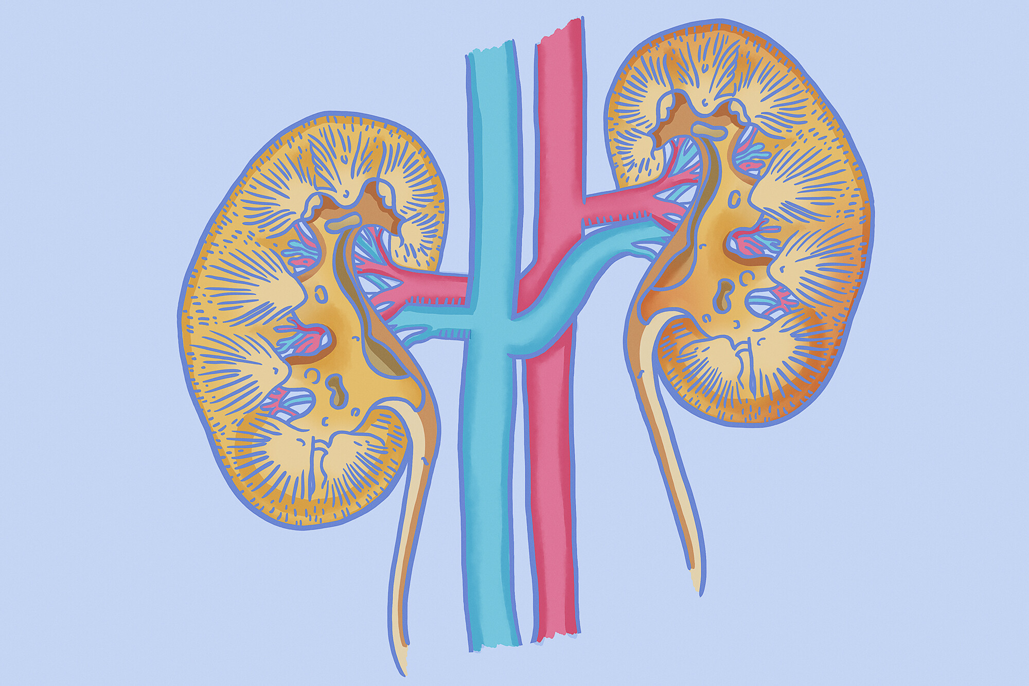
Penn Medicine researchers have built the first spatial map of the developing human kidney, showing that soluble signals from neighboring cells steer what each cell becomes.
A human kidney is built once, before birth, and the rules guiding its construction have been largely inferred from animal models. Researchers have now read those rules directly in human tissue. Experts at Penn Medicine have found that developing cells are steered by soluble signals released by their neighbors—and identified the insulin-like growth factor 2 (IGF2) as a key signal that sustains the kidney’s stem-like cells. The study is published in Nature Genetics and conducted by the Penn-CHOP Kidney Innovation Center, a research collaboration between the Perelman School of Medicine and Children’s Hospital of Philadelphia, working with bioengineers from Penn’s School of Engineering and Applied Science and the Institute for Regenerative Medicine.
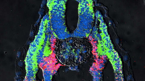
To capture development as it occurs in intact tissue, the team combined single-cell RNA sequencing with two spatial transcriptomics platforms, analyzing more than 700,000 cells from human fetal kidneys between 12.5 and 20.5 weeks of gestation. Conventional single-cell sequencing requires dissolving tissue into separate cells, which discards exactly what development depends on: where each cell sits and which signals reach it. The spatial approach preserves that context, letting the researchers place every cell, and every stage of its differentiation, back onto the map of the developing organ.