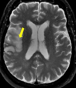
view more
Image source link: https://commons.wikimedia.org/w/index.php?title=Special%3AMediaSearch&type=image&search=FSE_T2_MRI_of_cerebral_infarction
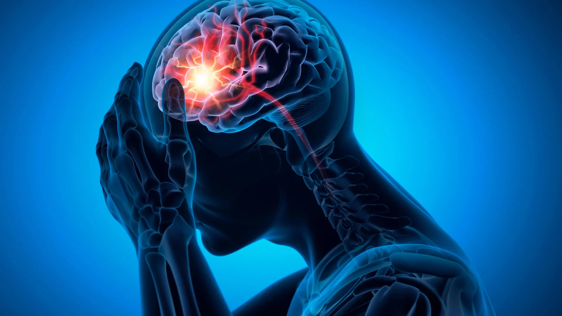
The buildup of fats, cholesterol, calcium, and immune cells within the blood vessel can lead to the narrowing or even complete blockage of the blood vessel. This condition, called stenosis, can lead to various disorders. Moyamoya disease (MMD) results from stenosis of the blood vessel in the neck that supplies blood to the brain (distal internal carotid artery (ICA)), as well as its branches. This leads to the suboptimal blood supply to the brain, a condition termed cerebral ischemia. Currently, the main treatment strategy for MMD involves the surgical augmentation of blood supply to the brain (neovascularization). However, neovascularization is associated with cerebral ischemia risks. The prediction of cerebral ischemic events after surgery has been challenging, mainly because the related risk factors have not been identified.
To address this knowledge gap, a research team led by Dr. Yuanli Zhao and Prof. Rong Wang examined the medical imaging features of 119 patients with ischemic MMD who underwent neovascularization surgery at the Department of Neurosurgery, Beijing Tiantan Hospital, Capital Medical University. The study was published in the Chinese Neurosurgical Journal on 14 April 2026. Commenting on the rationale of the study, Dr. Zhao remarked, “The incidence of cerebral ischemia after revascularization surgery in MMD is about 5%. The identification of risk factors for postoperative cerebral ischemia can aid in developing predictive tools and consequently improving patient outcomes.”