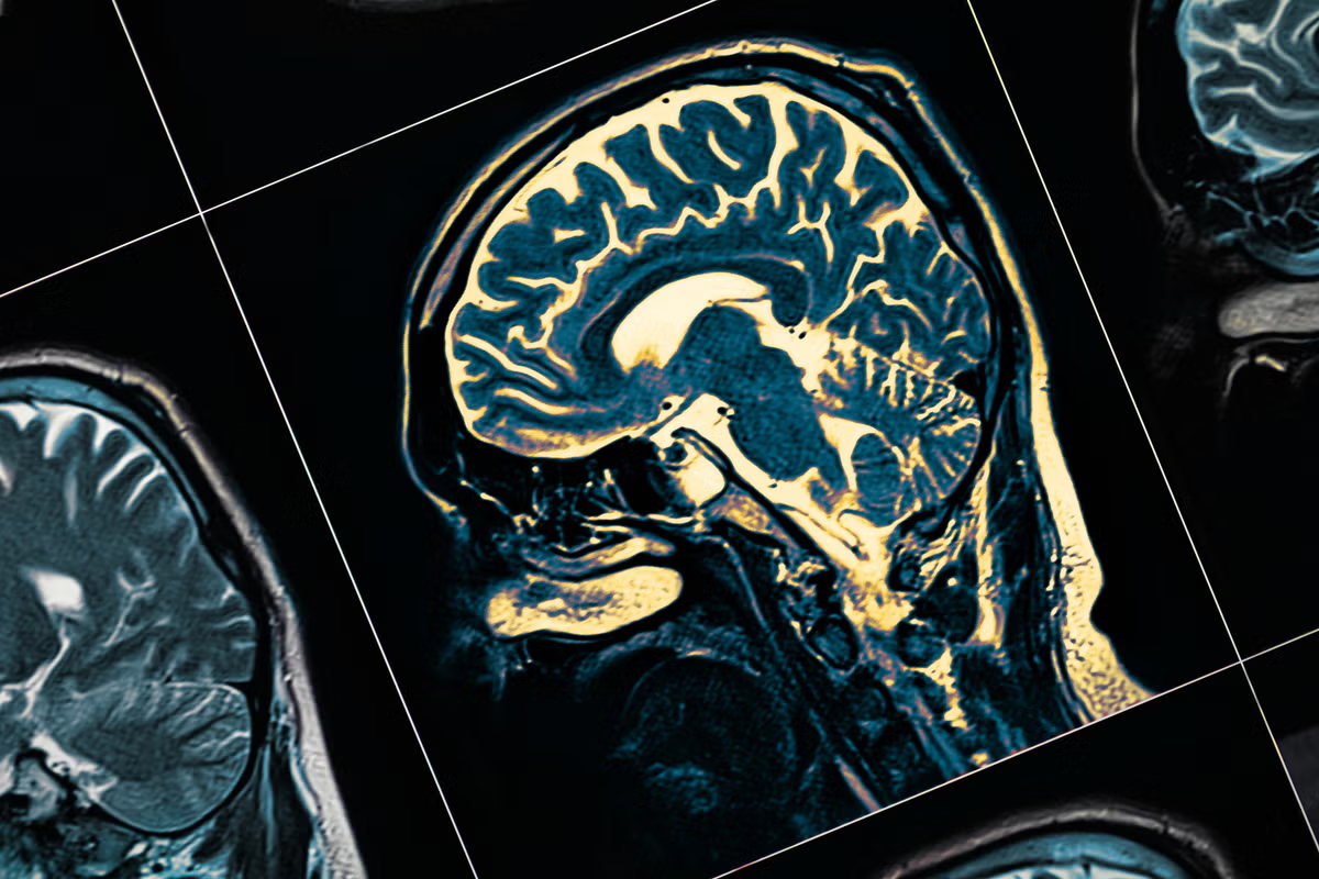
An EPFL-led study shows that individual brain connectivity patterns, unique to each person, can identify Parkinson’s patients with hallucinations and reveal early signs linked to cognitive vulnerability.Every brain is wired differently. Using functional MRI (fMRI), scientists can map how different brain regions functionally communicate with each other, creating a "connectome." These patterns are unique enough to act like brain fingerprints, opening new possibilities for personalized medicine.Parkinson's disease is best known for causing tremors and movement difficulties, but many patients also experience non-motor symptoms, such as hallucinations. The mildest form, known as minor hallucinations, includes fleeting sensations of a presence nearby or brief visual distortions. These symptoms affect up to 40% of patients, often appear early in the disease, and are linked to a higher risk of later cognitive decline.Most previous studies have compared groups of patients on average, potentially missing important differences between individuals. That matters in Parkinson's disease, where patients can follow very different neurological trajectories.Mapping Parkinson’s connectomeA team led by Sara Stampacchia and Olaf Blanke at EPFL's Laboratory of Cognitive Neuroscience has now applied brain fingerprinting to Parkinson's patients for the first time using resting-state fMRI, a widely used method for mapping brain connectivity with high spatial detail.Their work, published in Nature Mental Health, shows that these individual connectome signatures are preserved in Parkinson's disease and carry clinically meaningful information.The researchers worked with Parkinson's patients, with minor hallucinations and without. Each patient underwent a resting-state fMRI scan. Rather than asking patients to perform tasks, resting-state fMRI records the brain's spontaneous activity, capturing how different regions communicate with each other across the connectome.The team split each scan in half, treating the two halves as separate sessions, and tested whether each patient's brain connectivity pattern was consistent and unique enough to identify them from the group.Brain fingerprints reveal hidden differencesThe method correctly identified every patient from their brain connectivity pattern alone, regardless of whether they had hallucinations.But the networks driving those signatures differed between the two groups. When compared to patients without hallucinations, patients with minor hallucination showed a loss of distinctive features in the default mode network (DMN). Given that the DMN is among the most individually distinctive in cognitively healthy individuals, this reduction may constitute an early biomarker of the increased cognitive decline risk in this group.On the other hand, other networks – including cerebellar-cortical connectivity, the somatomotor and fronto-parietal networks - emerged as strong drivers of identifiability in this group. This is of interest, as somatosensory and motor integrations processes are thought to play a role in minor hallucinations pathogenesis, while fronto-parietal alterations have been linked to this symptom in previous work.Leveraging this uniqueness, the team proposed a new method to derive patient-specific networks and demonstrated that, in patients with minor hallucinations, the strength of connectivity in these networks were associated with scores on tests of frontal and executive cognitive function, even though most patients still had cognitive scores within the normal range overall.In patients without hallucinations, the same approach instead explained variability in motor symptom severity. This split suggests that the two groups may already be following different neurological trajectories, and collectively, these results show that is possible to characterise individualised brain alterations linked with clinical symptoms expression.The researchers also found that the brain fingerprint patterns aligned with different neurotransmitter systems in the two groups. In patients without hallucinations, the patterns were associated mainly with dopamine-related systems. In patients with hallucinations, they aligned more strongly with serotonin, GABA, and acetylcholine systems, all of which have previously been linked to hallucinations and cognitive decline in Parkinson’s disease.Toward earlier and more personalized careThe study shows that subtle, individual-level differences in brain function can be detected even at an early stage of disease, before major cognitive impairment becomes clinically apparent.The findings could help researchers better understand why some Parkinson's patients experience faster cognitive decline than others, and why different patients respond differently to treatments. However, the authors stress that the study involved a relatively small number of patients, and short scan acquisition times, and that larger studies with longer acquisitions will be needed before the approach could have clinical applications.Other contributorsUniversity of GenevaUniversity of BirminghamSant Pau HospitalUniversitat Autònoma de Barcelona (UAB)Centro de Investigación en Red-Enfermedades Neurodegenerativas (CIBERNED)Biomedical Research Institute (IIB-Sant Pau)Geneva University HospitalFundingSwiss National Science FoundationCARIGEST SA (Fondazione Teofilo Rossi di Montelera e di Premuda, and others)Parkinson SuisseEPFL Neuro X Post-doctoral Fellowship ProgramLeenaards FoundationSynapsis FoundationCIBERNED (Carlos III Institute)Italian Science Fund (FIS)Instituto de Salud Carlos III (ISCIII)PERISGeneralitat de CatalunyaReferencesSara Stampacchia, Fosco Bernasconi, Dimitri Van De Ville, Enrico Amico, Lada Kohoutová, Javier Pagonabarraga, Jaime Kulisevsky, Olaf Blanke. Patient-specific functional networks track early cognitive alterations and minor hallucinations in Parkinson’s disease. Nature Mental Health 08 June 2026. DOI: 10.1038/s44220-026-00657-x
Brain scans reveal early cognitive changes in Parkinson's
An EPFL-led study shows that individual brain connectivity patterns, unique to each person, can identify Parkinson’s patients with hallucinations and reveal early signs linked to cognitive vulnerability.
777 words~4 min read