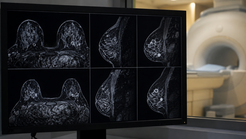
Enhanced, abbreviated MRI (AMRI) outperformed ultrasonography (US) as a screen for early liver cancer in high-risk patients with cirrhosis, according to a single-center, randomized clinical trial.
Overall, AMRI yielded significantly more early-to-advanced stage cancer among 759 patients: the per-patient detection rate in Barcelona-Clinic Liver Cancer (BCLC) stage 0, A, B, or C hepatocellular carcinoma (HCC) was significantly greater in those randomized to screening with hepatobiliary-phase image AMRI (HBP-AMRI) using gadoxetic acid than in those screened with US, at 8.5% versus 3.1% (P=0.002).
That screening performance gap widened in early-stage HCC (BCLC stage 0 or A), where per-person detection rates reached 7.7% in those screened with HBP-AMRI, compared with 2.9% of those screened with US (P=0.003). There was a difference even in detecting very-early BCLC stage 0 cases (6.1% vs 0.8%, P=0.001), reported Sung Won Chung, MD, of the University of Ulsan in Seoul, Korea, at the European Association for the Study of the Liver annual meeting in Barcelona.
"The bottom line is that HBP-AMRI significantly outperformed conventional ultrasound for HCC surveillance in high-risk patients with cirrhosis," Chung said during his presentation of the AMRIUS study. The results "support a risk-stratified surveillance approach."