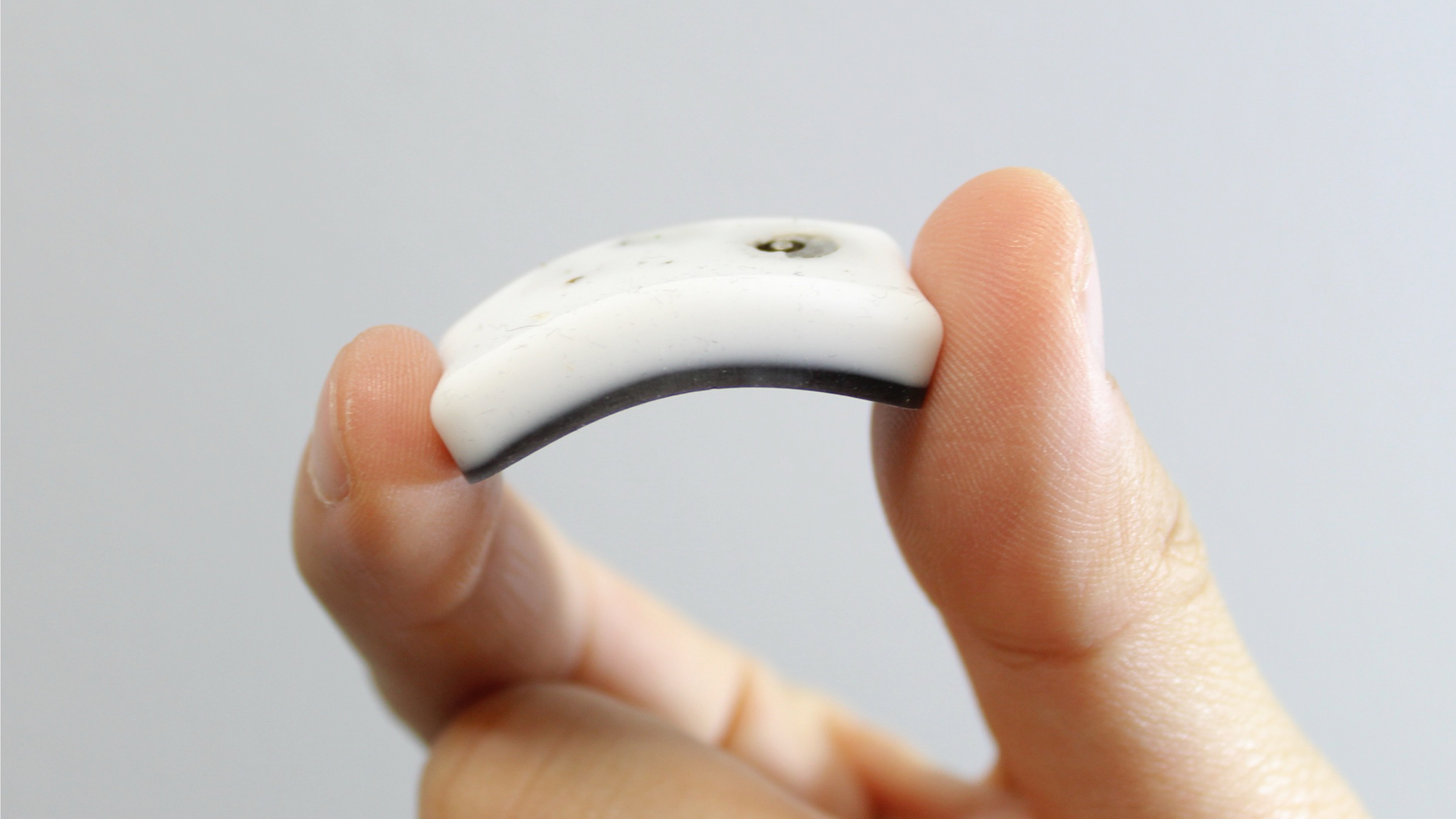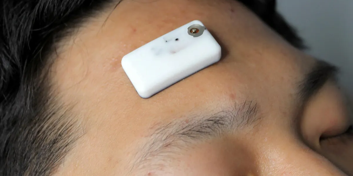
When you get a good night’s sleep, you aren’t just giving your brain a chance to rest. Sleep activates a system only discovered in 2012 that washes out brain waste. Called the glymphatic system, it’s comparable to the better-known lymphatic system that moves and filters fluids throughout your body. A healthy glymphatic system is linked to good cognitive function and could prevent neurodegenerative diseases like Alzheimer’s, but monitoring it during sleep has been practically impossible in humans, because today’s methods require noisy, confining MRIs and invasive spinal injections. A new wearable device developed by researchers at Georgia Tech and Seoul National University (SNU) could offer a safer and more sleep-friendly alternative. The technology shines near-infrared light to detect brain water, a soup of the fluids that constantly flood your brain. The brain water mixture contains cerebrospinal fluid (CSF), which is what the glymphatic system uses to flush out waste particles like plaques that block in-brain communication. Measuring total brain water could be a way to study how the glymphatic system moves CSF around to clean the brain, researchers say.The patch, the design of which was published this month in Science Advances, is intended for ease-of-use. It’s the size of a Band-Aid and less than a centimeter thick. Its soft silicone body conforms to the user’s forehead, and it doesn’t require a wired connection during sleep. Plus, it can be recharged and used over multiple nights, capturing more long-term information than what traditional sleep studies and MRIs can.“MRI is super expensive, it’s not really accessible, and more importantly, you cannot sleep under MRI imaging,” says W. Hong Yeo, Peterson professor in pediatric research at Georgia Tech. “With our device, we can naturally capture conventional sleep right at home.”What brain water could say about sleep and the glymphatic systemThe glymphatic system is essentially a network of tiny voids between veins, arteries, and cells that get flooded with CSF when brain cells relax. Sleeves called perivascular spaces, which surround blood vessels, deliver the fluid to the spaces between cells. Dr. Chang-Ho Yun, a professor of neurology at SNU’s Bundang Hospital, says that these intercellular spaces can expand by about sixty percent in sleeping mice, but observations of the change in humans remain indirect. “The human brain is densely packed with cells,” Yun says. “During sleep the alerting signal (noradrenaline) drops away, the cells shrink, and there’s room for cerebrospinal fluid to flow.” The new device uses a simple light trick to try to detect CSF changes. If you’ve ever held a flashlight to your palm in a dark room, you’ve seen the beam cause your hand to glow red. That’s because shorter wavelengths of light, like green and blue, get absorbed in your tissue. Red light breaks through and scatters back. The patch shines three different wavelengths of near-infrared light: two that are absorbed by blood cell proteins called hemoglobins, which deliver oxygen to the brain, and one that is absorbed by water. What’s scattered back gets picked up by the device’s photodetector. The pattern of absorption at each wavelength reveals how much blood and total brain water lie along the path of the light. Yun says that if total brain water rises while hemoglobin stays flat, the added water is not coming from blood, which could mean CSF is increasing and the glymphatic system is doing its job. This indirect measure is necessary because CSF does not reflect light all that differently from the other fluids in your brain.“Although it is indirect evidence, it’s compatible with known theory and known facts demonstrated in animals and humans,” Yun says. In his previous research, he found evidence that suggested glymphatic activity lowers during the REM stage of sleep. The study associated with the new patch, which measured sleep in four people, showed brain water measurements doing the same during the REM cycle. Still, he emphasizes that further research is necessary to determine if total brain water changes truly signal glymphatic activity.Lauren Hablitz, an assistant professor of translational neuromedicine at the University of Rochester, agrees that it’s hard to say whether the patch is actually monitoring the glymphatic system. Knowing for sure might even be impossible, she adds. “The brain is bathed in fluid, it sits in fluid, it floats in fluid,” she says. “Knowing whether it’s that pool of fluid or the perivascular space or the ventricles that’s changing is hard.”But Hablitz is optimistic about how the new tech can be used, even if it isn’t ultimately measuring the glymphatic system. She says that sleep research is “heavily focused” on electroencephalograms, or EEGs, which use electrodes placed on the scalp to measure the brain’s electrical activity. Yet most people with sleep issues have normal EEG readings, she says.“Maybe something like this patch, that can look at another aspect of the biology that isn’t just neuronal activity, can start saying something about what’s actually happening in sleep disruption,” she says.
Tiny Sleep Patch Could Track Brain's Nightly Cleansing Ritual
Monitoring the elusive activity of the glymphatic system could aid Alzheimer’s research
815 words~4 min read