by Sara Pecchia
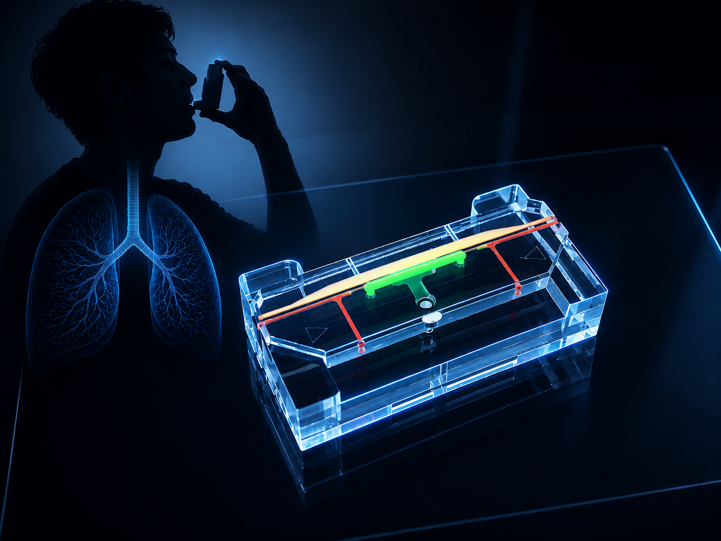
Carnegie Mellon University researchers have co-created a miniature airway organoid model that more accurately reflects real lung biology by pairing lung-derived biomaterials with an “apical-out” design, allowing scientists to better study infections, environmental exposures, and respiratory disease.
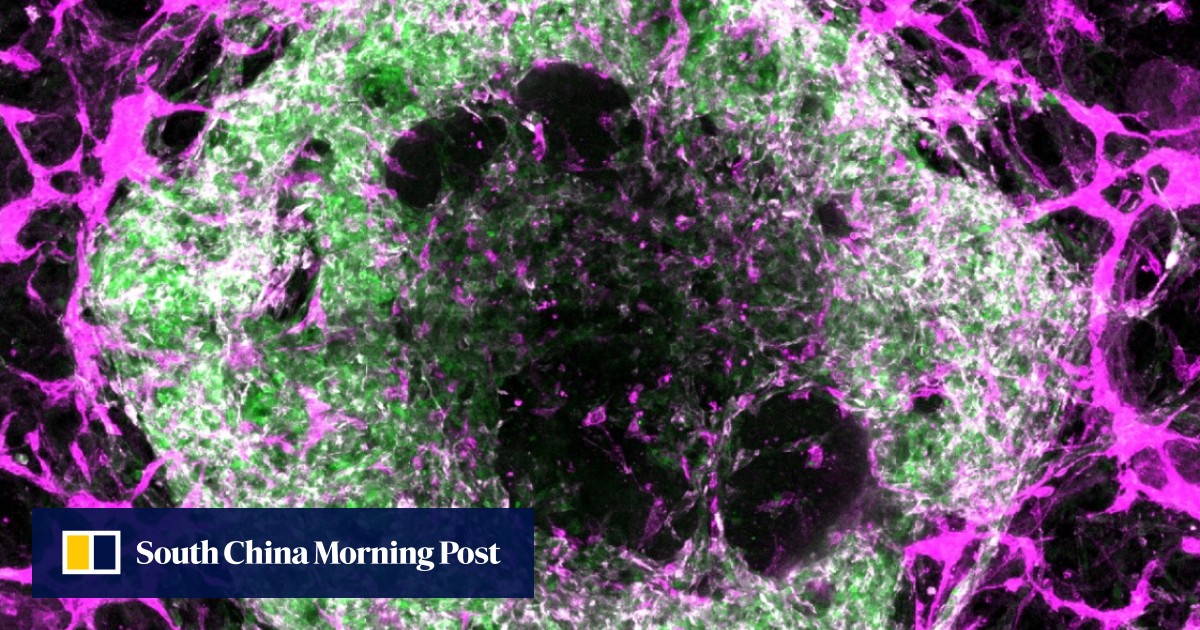
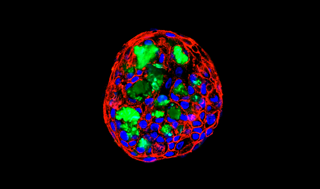
Researchers from Carnegie Mellon University’s Ren lab (Engineered Morphogenesis Group) have co-created a miniature model of the human airway that behaves more like real lung tissue. By combining natural lung-derived biomaterials with a special “apical-out” organoid design, the team created a system that captures two key features of airway biology that existing models rarely achieve together.
The lung forms one of the body’s most important interfaces with the outside world. With every breath, airway tissues encounter airborne particles and pathogens. Understanding how airway cells respond to these exposures is essential for studying diseases such as respiratory infection, asthma, and chronic obstructive pulmonary disease.
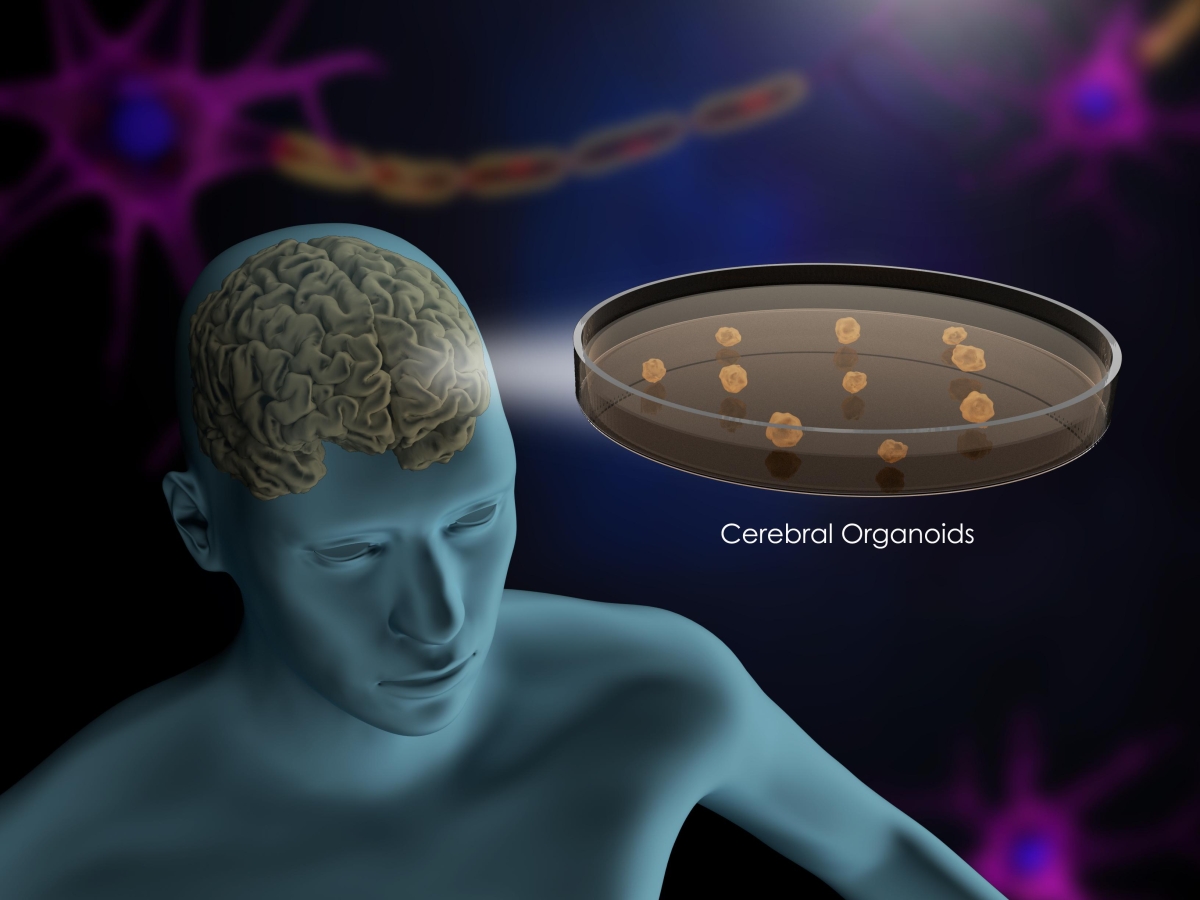
Scientists often grow miniature tissue structures known as organoids to model human organs. These three-dimensional cultures reproduce some features of real tissue. However, many airway organoid systems capture only part of the biological environment.