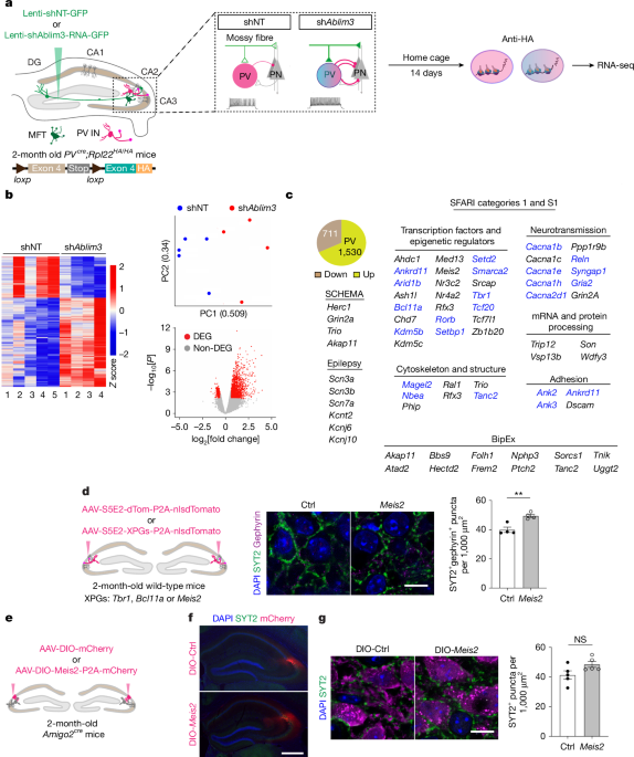
MainThe hippocampus has a crucial role in memory formation, storage and retrieval to facilitate the experience-dependent calibration of motivated and defensive behaviours2. During the early postnatal period, experience refines the hippocampal circuitry to influence cognitive trajectory20. Disruptions in hippocampal circuitry result in maladaptive neural circuit functions, cognitive impairments and seizures that characterize different NDDs, including autism spectrum disorder (ASD). The identification of ultrahigh-confidence genetic risk factors for NDDs underscores the need to instantiate how experience and genetic risk interact to impair experience-dependent mechanisms that support hippocampal-dependent cognition.PV INs in the dentate gyrus (DG)–CA3/CA2 circuit of the hippocampus influence encoding, storage, retrieval and routing of memories through activity-dependent regulation of CA3/CA2 PN perisomatic inhibition4,5,6,7,9,10,11. Feedforward inhibition of CA3/CA2 PNs mediated by PV INs dictates their spiking, synchronization of PN activity to form ensembles and the generation of network oscillations to regulate intrahippocampal and inter-regional communication in hippocampal–cortical–subcortical networks11,21,22. To exert these effects on circuitry and network properties, PV INs cell-autonomously coordinate experience-dependent changes in their intrinsic properties, structural reorganization of axonal arborizations and perisomatic synapses in CA3/CA2, regulation of feedforward inhibition of CA3/CA2 and synaptic plasticity9,10,12,13,14,15. Here, these properties are collectively referred to as experience-dependent PV IN plasticity. Loss of PV IN functions in NDDs, including ASD, bipolar disorder, schizophrenia and epilepsies, may arise from genetic risk factors that impair experience-dependent refinement of inhibition mediated by PV INs during the early postnatal period16,17,18,23,24.Transcription factors and epigenetic regulators co-ordinate changes in gene expression that underlie synaptic physiology, synaptic and structural plasticity and input–output connectivity to mediate experience-dependent inhibitory and excitatory neuron plasticity. We know a substantial amount about the molecular mechanisms that regulate cortical PV IN identity and experience-dependent plasticity12,17,25,26,27, but less about developmental regulators of hippocampal PV IN properties13,28,29. Moreover, evidence for transcription factors and epigenetic regulators that control experience-dependent PV IN plasticity in the adult hippocampus is scarce.PV INs in CA3/CA2 retain experience-dependent plasticity in adulthood. Specifically, learning6,10 and social experience9 increase mossy fibre excitatory synaptic inputs onto PV INs to trigger experience-dependent PV IN plasticity. This process subsequently leads to increased feedforward inhibition in DG–CA3/CA2 circuits9. Enhanced mossy fibre excitatory drive onto PV INs increases intrinsic excitability, inhibitory synapses onto CA3/CA2 and perisomatic inhibition of CA3/CA2 PNs to support spatial and social memory9,10. At a network level, DG recruitment of PV-IN-mediated inhibition of CA3/CA2 PNs promotes stable and context-specific neuronal ensembles in hippocampal–cortical networks and network oscillations during memory consolidation10,11. Here we build on this previous work to screen for cell-autonomous regulators of XPGs in the adult CA3/CA2 circuit. We report a suite of candidate XPGs, including transcription factors and epigenetic regulators, that are encoded by ultrahigh-confidence risk genes for NDDs and epilepsies. We test the hypothesis that restoration of experience-dependent PV IN plasticity in the adult hippocampus in a widely used NDD risk mouse model is sufficient to reverse developmental deficits in circuitry, ensemble specificity, sharp-wave ripple (SWR) properties, seizures and cognition.Discovery of XPGs in CA3/CA2To identify XPGs in CA3/CA2 PV INs, we designed an in vivo functional screen in adult mice to isolate translated mRNAs from CA3/CA2 PV INs that receive increased mossy fibre inputs, a trigger for experience-dependent PV IN plasticity (Fig. 1a). To this end, we used lentiviruses to express short hairpin RNA (shRNA) to downregulate Ablim3 (shAblim3) or a non-target shRNA as a control (shNT). ABLIM3 is a molecular brake of connectivity between mossy fibre filopodia and PV INs, and its downregulation in DG mossy fibres9,10,11 reproduces the effects of experience on mossy fibre filopodial inputs onto PV INs9,10,11. We used PVcre;Rpl22HA/HA mice30 (PV is also known as Pvalb), in which expression of the haemagglutinin (HA) epitope-tagged ribosomal protein L22 (RPL22) is restricted to PV INs10. After optimization of the signal-to-noise ratio for mRNA isolation from this extremely sparse population of PV INs31 (Extended Data Fig. 1a,b), we compared mRNAs biochemically isolated from genetically tagged PV INs in the naive state versus an experience-induced activated state in CA3/CA2. Using this approach, we identified differentially expressed candidate XPGs associated with the PV IN activated state (Fig. 1b,c and Supplementary Table 1). Significantly upregulated genes included 82 category 1 and S1 ASD genes reported in the SFARI database (https://gene.sfari.org/)32. Other genes identified included 4 genes causally implicated in schizophrenia through exome sequencing (data from the Schizophrenia Exome Sequencing Meta-analysis (SCHEMA) project)33 and 12 bipolar disorder candidate genes, including AKAP11 (P < 0.01, out of a total of 64 bipolar disorder candidate genes that scored P < 0.05 according to Bipolar Exome (BipEx) data; https://bipex.broadinstitute.org/)34. A curated and updated annotated XPG list is available online (https://xpg-atlas.onrender.com/). By contrast, we identified only three category 1, syndromic ASD genes in the list of significantly downregulated XPGs (Supplementary Table 1). Therefore, we focused on upregulated XPGs in this study. A third of the identified ASD genes encode histone methyltransferase, histone demethylases, chromatin remodelling factors and transcription factors, the functions of which in PV INs are poorly defined (Supplementary Table 1). Gene set enrichment analysis of all the genes upregulated in the experience-induced PV IN activated state suggested that these XPGs have a role in synaptic physiology, structural remodelling and presynaptic terminal specialization (Extended Data Fig. 1c). Half of the ASD-linked upregulated XPGs are implicated in epilepsies, consistent with the high incidence of seizures in individuals with ASD (https://gene.sfari.org/) (Fig. 1c). Moreover, individuals with ASD or schizophrenia often have loss-of-function mutations in these genes, and analyses of data from BipEx suggest that the bipolar disorder risk candidate XPGs have damaging missense or protein-truncating mutations. Because the transcripts of these XPGs are upregulated or poised for translation in PV INs in response to increased mossy fibre inputs, our findings indicate that loss of PV IN experience-dependent plasticity is a convergent mechanism for different NDDs, including ASD, bipolar disorder, schizophrenia and epilepsies.Fig. 1: Discovery of XPGs in adult CA2/CA3.a, Biochemical isolation and translatome sequencing of naive-induced and mossy-fibre-triggered (MFT) activated PV INs in adult PVcre;Rpl22HAf/f mice. Lentiviruses expressing shAblim3–GFP or shNT–GFP were injected into DG, and CA2/CA3 regions were microdissected 14 days later. n = 6 mice (3 male and 3 female) per sample, 5 samples, total 30 mice for shNT; 4 samples, total 24 mice for shAblim3. b, Left, heatmap of expression levels for differentially expressed genes (DEGs), shown as normalized z scores relative to the average expression of a given gene across all samples. DEGs were identified on the basis of the false discovery rate (FDR) adjusted for multiple hypothesis testing and estimated using EdgeR (fold change > 1.5 and FDR < 0.05). Right, top, principal component analysis plot of PV IN translatomes. The first two principal components (PC1 and PC2) are shown with corresponding fractions of variance. Bottom, volcano plot showing significance (–log10[P]) versus the magnitude of change (log2[fold change]) of gene expression. DEGs are indicated in red. c, Top left, pie chart showing the numbers of upregulated and downregulated XPGs. Bottom left and right, upregulated XPGs linked to ASD, bipolar disorder and schizophrenia are listed. XPGs in blue are also implicated in epilepsies. d, Left, experimental design. S5E2-driven AAVs expressing control (dTomato) or Meis2 were injected into CA2/CA3 of wild-type mice. Right, representative images and quantification of SYT2+gephyrin+ puncta density in CA3 (n = 4 mice per group, P = 0.0066). e–g, Experimental design. AAVs expressing DIO-mCherry (DIO-Ctrl) or DIO-Meis2-mCherry (DIO-Meis2) were injected into CA2 of Amigo2cre mice (e); and representative images for CA2-specific viral expression (f), SYT2+ puncta and quantification in CA2 (g) are shown (n = 5 mice per group). Scale bars, 10 μm (d,g) and 500 μm (f). NS, not significant; **P < 0.01 using two-tailed unpaired t-test with Welch’s correction (d,g). All experiments were performed in male and female mice, data are mean ± s.e.m.The upregulation of select candidate ASD-linked XPGs (Meis2, Bcl11a and Tbr1; https://gene.sfari.org/) and the SCHEMA gene Herc1 (ref. 33) in activated CA3/CA2 PV INs was validated by qPCR (Extended Data Fig. 1d). Therefore, we asked whether inducing the expression of these XPGs in PV INs via S5E2 enhancer-driven AAV vectors35 promotes the formation of PV IN–CA3/CA2 synapses in adult mice. Virus-mediated expression of Tbr1, Bcl11a, Meis2 or Herc1 in S5E2-targeted PV INs increased PV IN synapses in CA3/CA2 PNs (Fig. 1d and Extended Data Fig. 1f–h). Meis2 is expressed at negligibly low levels in a subpopulation of hippocampal PV INs in the hippocampus36, where its function is unclear. MEIS2 is a homeodomain-containing transcription factor that acts as a cellular-context-dependent coactivator to regulate the developmental specification of GABAergic projection neurons19,37 and maintains intrinsic excitability and terminal arborization in sensory neurons38. On the basis of these observations, we prioritized Meis2 as a candidate XPG to restore experience-dependent PV IN plasticity in NDDs. As the S5E2 enhancer preferentially, but not exclusively, targets PV INs in CA3/CA2 (Extended Data Fig. 1e), we asked whether Meis2 overexpression in CA2 PNs may also contribute to the increase in PV IN synapses onto CA2 PNs. Using a CA2-specific Amigo2cre mouse line in combination with an AAV-DIO-Meis2-P2A-mCherry virus, we found that Meis2 overexpression in CA2 PNs does not increase PV IN synapses onto mCherry+ CA2 PNs (Fig. 1e–g and Extended Data Fig. 1i). Many NDDs, including ASD, are characterized by a range of developmental deficits, intellectual disability, seizures and impaired social cognition. Therefore, we selected Cntnap2 knockout (Cntnap2−/−) mice to test our hypothesis. The goal was not to model a specific NDD but to use an NDD risk mouse model, like Cntnap2−/− mice, which exhibits a range of developmental deficits, including altered neuronal migration and excitability, loss of PV INs, cognitive impairments and seizures by around 6 months of age39,40,41,42.
Procognitive restoration of PV neuron plasticity in neurodevelopmental disorders - Nature
Expression of Meis2 in hippocampal parvalbumin-expressing interneurons in an adult mouse model of neurodevelopmental disorders rescues experience-dependent plasticity, improves memory and network function and suppresses seizures.
18,295 words~83 min read