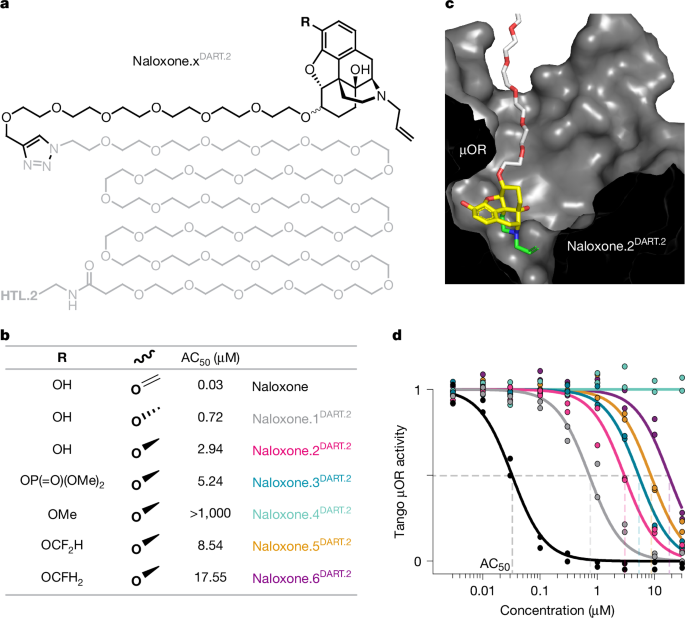
MainOpioids remain essential analgesics for managing acute pain following injury or surgery. Although they are generally effective, around 2% of opioid-naive individuals transition to extended opioid use26,27—a minority that nonetheless accounts for around 75% of opioid use disorder cases in the USA28. Opioids signal to multiple brain cell types, yet it is unclear whether their multifaceted short-term effects can be separated from the cumulative neural-circuit changes that drive abuse progression. Diverse synthetic opioid receptor agonists have been developed, yet none dissociate analgesia from reward learning—a process that is implicated in early stages of abuse progression1,2,3. Aiming to achieve this dissociation, adjuncts that disrupt opioid-mediated dopamine signalling are being developed29,30,31, capitalizing on the central role of dopamine in reward learning4. However, dopamine is also essential for the modulation of central pain perception by opioids5,6,7,8,9,10,11,12,13, underscoring the need for alternative approaches.Against this backdrop, a recent double-blind placebo-controlled human trial tested the pro-cholinergic drug galantamine, motivated by a neocortical cognitive hypothesis, and reported significantly reduced illicit opioid use18. Genetic variants of acetylcholinesterase that accelerate acetylcholine breakdown have also been associated with human vulnerability to heroin addiction19, further supporting the idea that sustained cholinergic tone may be protective. Moreover, pro-cholinergic drugs attenuate opioid-reward learning in rodent models20,21,22,23, suggesting that cholinergic tone may influence opioid-reward processing across species. Nevertheless, the relevant circuits remain unresolved. Cortical cholinergic architectures are poorly conserved across mammals32 and galantamine did not improve cognitive outcomes in the human trial, challenging a purely neocortical explanation33,34.The nucleus accumbens (NAc) is a subcortical circuit that is implicated in central pain perception and reward learning35,36, with substantially higher cross-species conservation37—making it an attractive candidate for a conserved cholinergic influence on opioid-reward learning. The NAc is a major target of opioid-evoked dopamine release, triggered primarily by μ-opioid receptor (μOR)-mediated disinhibition in the distant ventral tegmental area38. By contrast, local cholinergic interneurons (CINs) are the primary source of acetylcholine in the NAc39. Local opioid manipulations in the NAc can modulate opioid reinforcement40,41,42,43, but prior work has not tested whether NAc opioid receptors are required for the acquisition of associative opioid-reward learning or identified the cell types involved. Specifically, efforts to establish a causal role for CINs have been inconclusive. Targeted ablation of CINs leads to dopamine hypersensitivity and broad circuit adaptations44,45,46, whereas CIN-specific opioid receptor knockouts had little effect on the acute or cumulative effects of morphine47. Both approaches are susceptible to developmental and network-level compensation, leaving the question of whether opioid signalling on CINs contributes to the acquisition of opioid-reward learning unresolved. Addressing this question requires tools that can acutely block native opioid receptors on CINs with cellular specificity.Here, we address this challenge using the DART (drug acutely restricted by tethering) platform14,15 to develop naloxoneDART—a cell-type-specific variant of the opioid receptor antagonist naloxone, preserving the temporal precision and molecular specificity of traditional pharmacology. CIN-specific naloxoneDART abolished the acquisition of morphine conditioned place preference (CPP) in a target-engagement-dependent manner. This effect was not attributable to nonspecific contextual learning or motor deficits: saline habituation was enhanced between sessions and unchanged within sessions, whereas morphine-evoked hyperlocomotion, locomotor sensitization, and acute analgesia remained intact. Neurochemical measurements further revealed that CIN-specific naloxoneDART prevented morphine-induced acetylcholine reductions without measurably altering dopamine increases in the NAc. Collectively, these findings identify a CIN-dependent cholinergic gate that can decouple opioid-evoked accumbal increases in dopamine from associative reward learning.Developing a cell-type-specific opioid antagonistBuilding on a complementary study that establishes the feasibility of tethered opioid receptor antagonism48, we developed naloxone.xDART.2, a cell-type-specific version of naloxone using the second-generation DART.2 platform14,15, which enables thousandfold cellular specificity, provides matched control and tracer reagents, and offers a quantitative framework to bound tethered versus ambient ligand exposure during behavioural assays.DART uses an adeno-associated virus (AAV) to achieve cell-type-specific expression of the HaloTag protein (HTP). This protein enables selective tethering of DART ligands, comprising the drug of interest conjugated to the HaloTag ligand (HTL). Within minutes of application, DART ligands accumulate on targeted cells, generating cell surface concentrations orders of magnitude above ambient levels. HTL forms a covalent bond to HTP, maintaining tethered drug on the cell surface for days14,15.Using naloxone as a scaffold, we synthesized six naloxone.xDART.2 variants, differing in their C6 linker-attachment stereochemistry and C3 phenol-group substitution (Fig. 1a–c). All variants contained an identical polyethylene glycol (PEG36) linker and second-generation HTL.2 capture moiety15. For each variant, we determined AC50—the ambient concentration needed for 50% pharmacological effect—on 3 opioid receptor subtypes (μOR, δ-opioid receptor (δOR) and κ-opioid receptor (κOR); Fig. 1d and Extended Data Fig. 1). Four out of the six naloxone.xDART.2 variants were suitably attenuated to minimize ambient-drug effects (AC50 > 1 µM) while preserving efficacy at the high concentrations achieved by tethering14,15. We focused on naloxone.2DART.2 (hereafter naloxoneDART), which features an AC50 of approximately 3 µM against the μOR and comparable potency against the δOR and κOR (Fig. 1d and Extended Data Fig. 1).Fig. 1: Development of naloxoneDART.a,b, Chemical structure of naloxone.xDART.2 variants. a, The naloxone.xDART.2 backbone. b, Substituents and associated AC50 values for naloxone.xDART.2 variants. c, Naloxone.2DART.2 docked to μOR (Protein Data Bank (PDB): 4DKL). d, Dose–response curves showing Tango μOR activity versus naloxone.xDART.2 concentration (colours as in b). Data were normalized to the minimum and maximum of naloxone response from each of two replicate plates; lines are Langmuir fits used to derive AC50 for each ligand (values in b).Source dataNaloxoneDART makes CINs insensitive to morphineWe next validated these reagents using CIN pacemaker activity as a functional readout. To control for ambient naloxoneDART exposure, we developed a control condition (controlDART) containing an equimolar mixture of naloxone.2PEG (lacking HTL) and blankDART.2 (lacking naloxone). Critically, controlDART and naloxoneDART conditions have the same amount of PEG-linked naloxone, so any ambient effects are matched across conditions (Fig. 2a). The HTP-tethering process is also matched owing to equivalent amounts of PEG-linked HTL, leaving the presence or absence of a physical linkage between naloxone and HTL as the key experimental variable (Fig. 2a). Finally, both conditions were supplemented with a small amount of the fluorescent tracer Alexa647DART.2, comprising 9% of the tetherable compound (Fig. 2a). This tracer has been shown to exhibit comparable rates of diffusion and capture to other DART ligands, ensuring that the ratio of ligands remains stable during the delivery and tethering process14,15.Fig. 2: Morphine control of CIN pacemaking is blocked by tethered naloxoneDART.a, Schematic of ligand conditions: naloxoneDART contains a 1:10 ratio of Alexa647DART.2 and naloxone.2DART.2; controlDART replaces naloxone.2DART.2 with an equimolar mixture of naloxone.2PEG and blankDART.2. b, Acute brain slice incubated in naloxoneDART for 15 min. Right, single-plane optical section (confocal, 63× objective) shows cytosolic ChAT immunostaining, nuclear NLS–Tomato marking AAV expression, and surface Alexa647DART. Representative image from one of three cells from one mouse with similar results. Scale bar, 10 μm. c, Example pacemaker firing of a CIN pretreated with controlDART, showing the first 3 min of 1 µM morphine application. d, Example pacemaker firing of CIN pretreated with naloxoneDART (format as in c). e, Top, mean untreated CIN pacemaker firing in response to 1 µM morphine. Grey shading defines pre- and post-morphine intervals. Waveform shows baseline-normalized firing rate (FR). Data are mean ± s.e.m. over 10 cells. Bottom, summary of pre- and post-morphine CIN pacemaker firing rate (FR). Each connected symbol pair represents one cell. Data are mean ± s.e.m. over 10 cells. Two-way repeated-measures ANOVA across all data from e–g (bottom), revealing a significant treatment × time interaction (F4,39 = 8.30, P = 6.06 × 10⁻⁵). Tukey test showed a significant pre- versus post-morphine difference in untreated cells (P = 1.5 × 10⁻⁷). f, Top, mean CIN pacemaker firing response to 1 µM morphine with controlDART pretreatment. Data are mean ± s.e.m. over 7 cells. Bottom, Tukey test shows a significant difference between pre- and post-morphine in controlDART cells (P = 0.0005; format as in e). Data are mean ± s.e.m. g, Top, mean CIN pacemaker firing response to indicated morphine concentration with naloxoneDART pretreatment. Bottom, Tukey test shows that cells pretreated with naloxoneDART were unaffected by 1 µM morphine (P = 0.54, n = 7 cells), 10 µM morphine (P = 0.85, n = 11 cells) or 100 µM morphine (P = 0.75; n = 9 cells). Data are mean ± s.e.m. *P < 0.05, **P < 0.01, ***P < 0.001, ****P < 0.0001; NS, not significant (P ≥ 0.05).Source dataWe prepared acute coronal brain slices from ChAT::Cre mice, which were made to express HTP in accumbal CINs with an optimized viral strategy (Extended Data Fig. 2). Following 15 min incubation with naloxoneDART or controlDART, tethered ligand was apparent on the cell surface, with negligible intracellular penetration (Fig. 2b). Thereafter, whole-cell recordings measured intrinsic CIN pacemaking at baseline and following the application of morphine (Fig. 2c,d). Consistent with previous reports49, 1 μM morphine eliminated pacemaking in untreated slices (Fig. 2e) and in slices preincubated with controlDART (Fig. 2c,f). By contrast, CINs pretreated with naloxoneDART became morphine-insensitive, maintaining baseline pacemaking even in the presence of 100 μM morphine (Fig. 2d,g).Notably, morphine is cell-permeable and can reportedly activate intracellular opioid receptors50, which are inaccessible to cell-impermeable reagents such as naloxoneDART. Thus, these data indicate that morphine regulates CIN pacemaking primarily via surface receptors, consistent with prior evidence for specialized plasma membrane signalling51.Chemical and spatial specificity of naloxoneDART
A cholinergic hub in the nucleus accumbens gates opioid-reward learning - Nature
Cell-type-specific opioid blockade in accumbal cholinergic interneurons abolishes morphine-reward learning without impairing analgesia and decouples morphine-evoked dopamine elevations from acetylcholine dips, suggesting pro-cholinergic strategies to limit early opioid reward acquisition.
15,189 words~69 min read