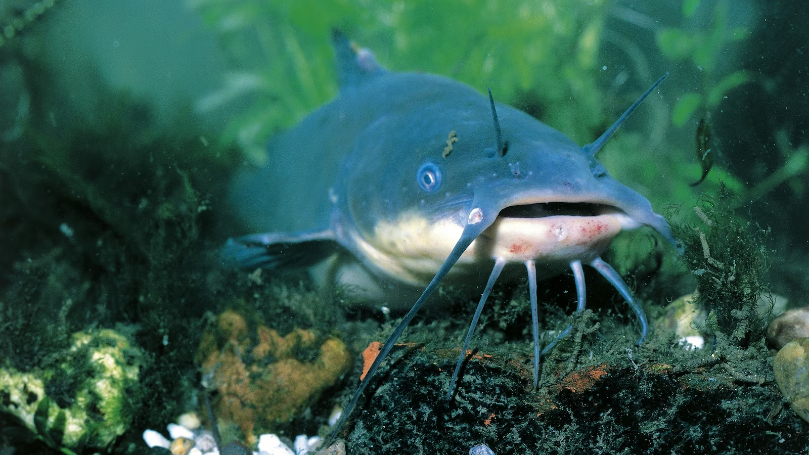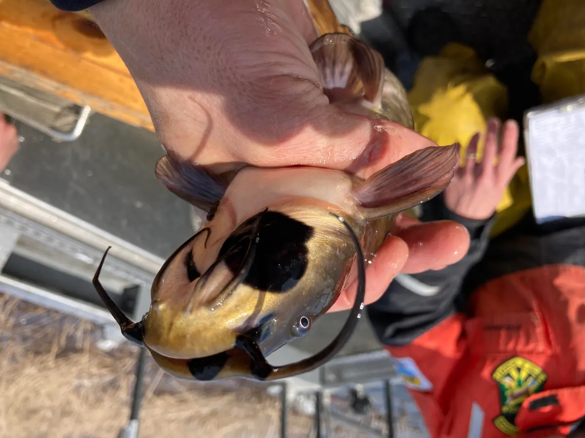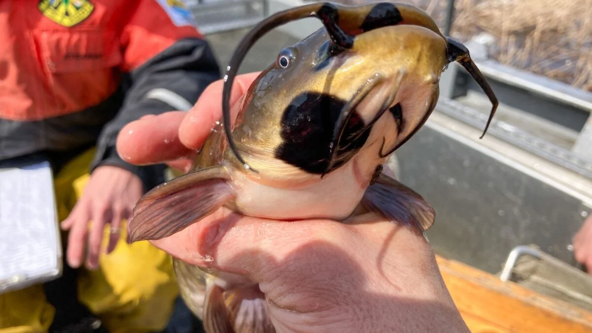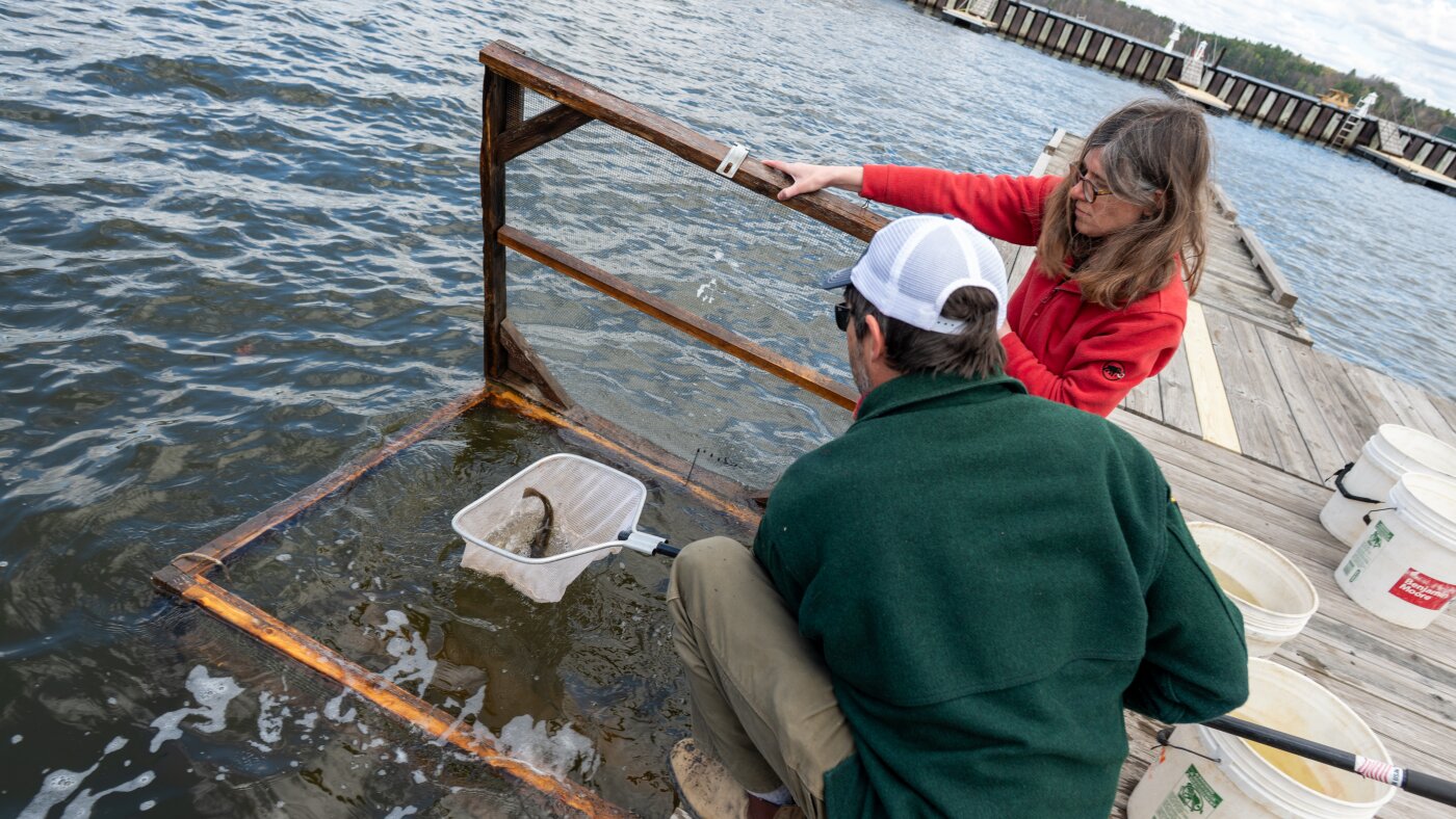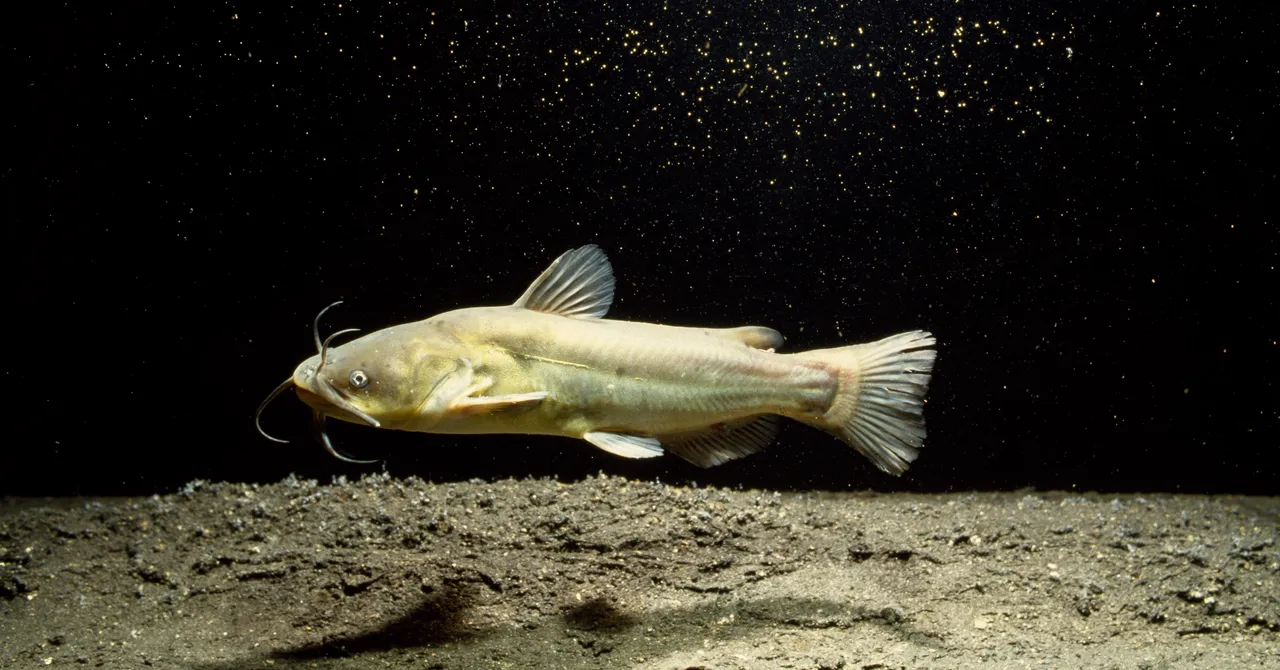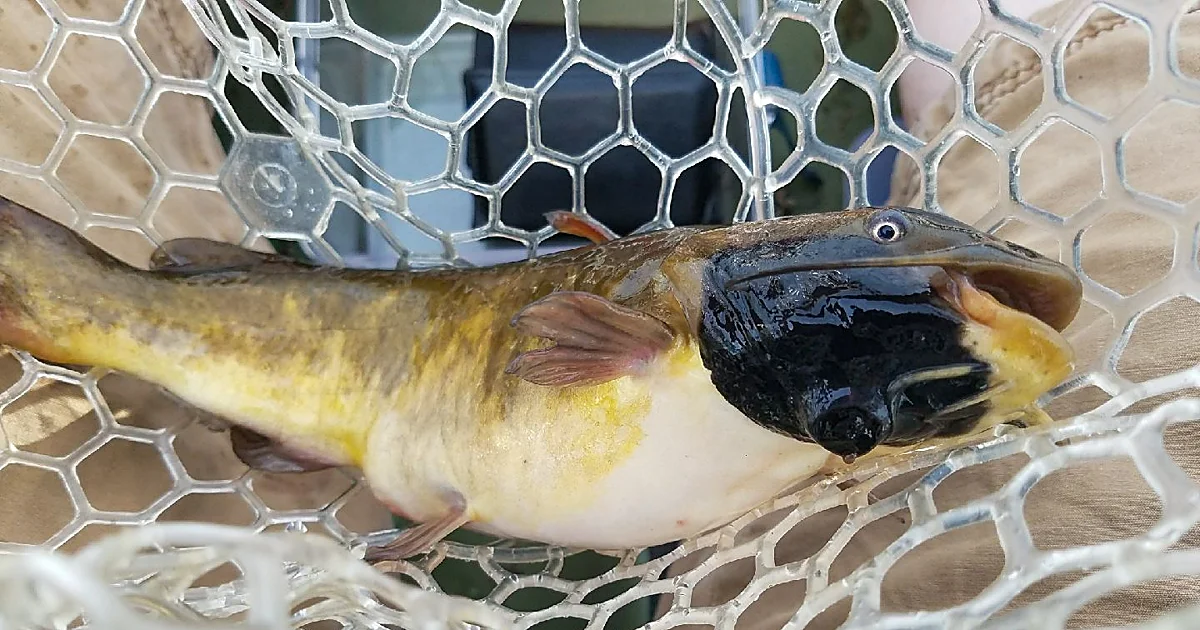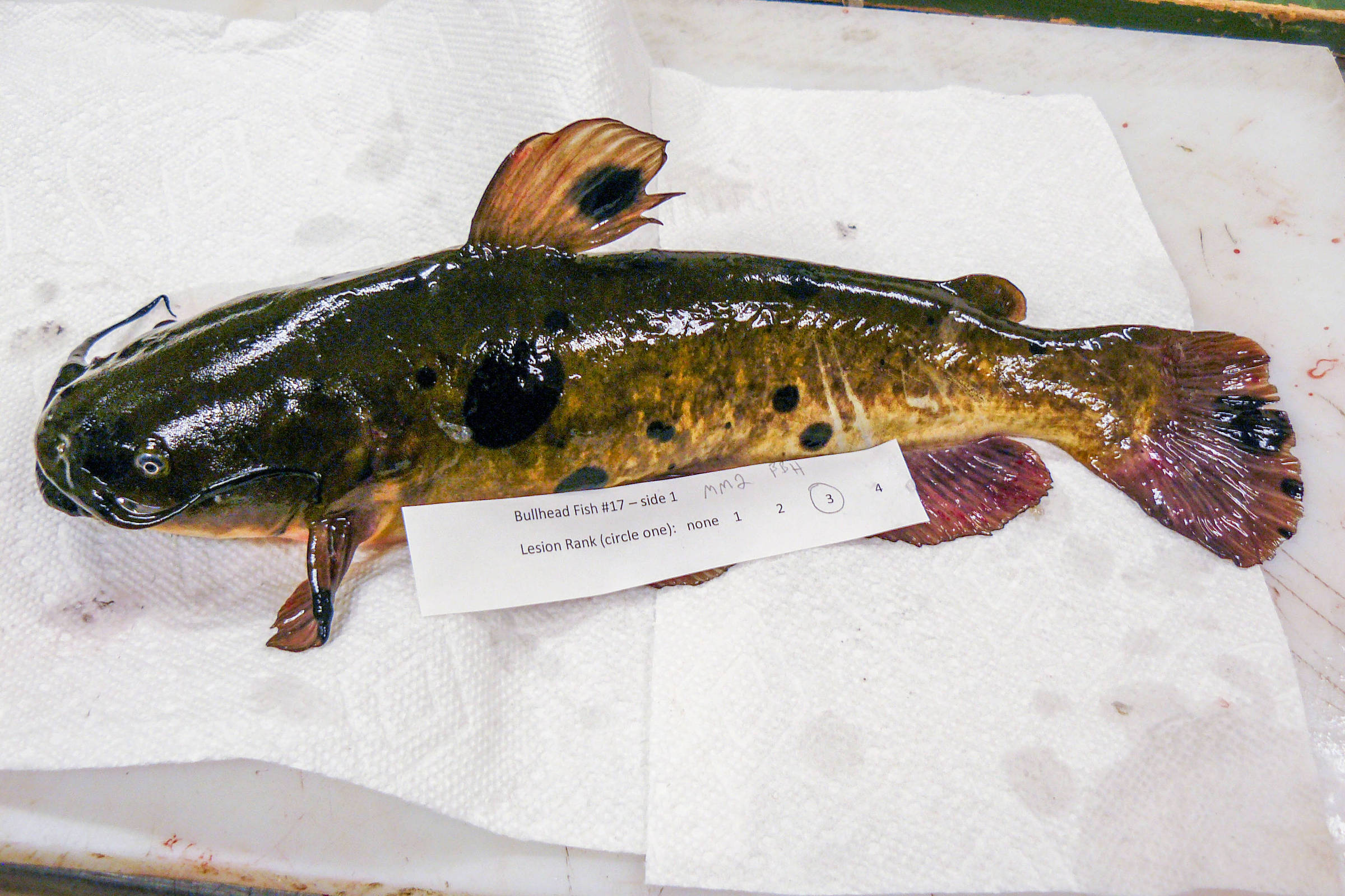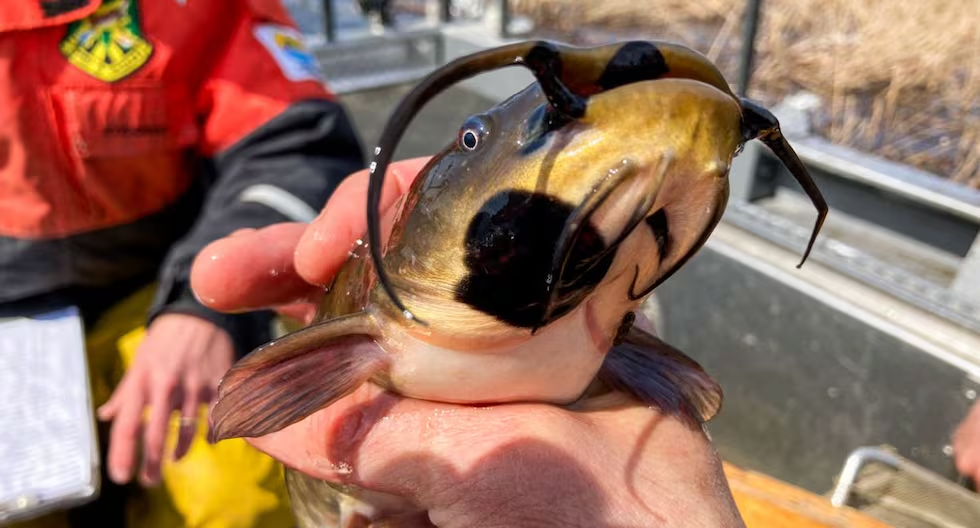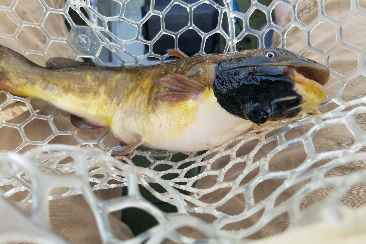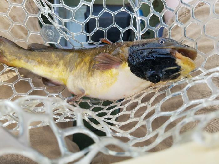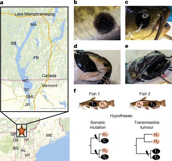
MainIn 2012, anglers and biologists began observing a high proportion of brown bullhead (A. nebulosus, a type of catfish) with raised black lesions in Lake Memphremagog, a freshwater lake in Vermont, USA, and Quebec, Canada1 (Fig. 1). A study determined these raised black lesions to be malignant melanomas, on the basis of histopathological analysis, and observed these lesions in 23–37% of brown bullhead sampled between 2014 and 2017 (ref. 1). Melanomas have been reported from brown bullheads in other bodies of water, but generally at considerably lower incidence1. Brown bullheads are widely distributed across Eastern North America and are considered an indicator species of water quality14. The high incidence of cancer in Lake Memphremagog could indicate a local cancer-causing contagion or contaminant. One hypothesis is that extensive flooding in 2011 from tropical storm Irene led to reduced water quality of the lake due to runoff pollution. Monitoring changes to water quality is of great concern to biologists and the public, as this lake is the source of drinking water for more than 175,000 people15.Fig. 1: Brown bullhead sampling, example melanistic lesions and hypotheses of tumorigenesis.a, The locations for field collection in Lake Memphremagog: Fitch Bay (FB), Magog Area (MA), Hospital Cove (HC; also designated North Main Lake east and west), Sargent’s Bay (SB), Scott’s Cove (SC), South Bay/Johns River (JR) and South Main Lake (SML). The locations for field collection of reference individuals: Sandy Point (SP) and Larabee’s (LB) in Lake Champlain, Lake Bomoseen (BS), Hoyts Landing (HL) in the Connecticut River, Ticklenaked Pond (TP), Village Pond (VP) and Hermon Pond (ME). b–e, Examples of melanistic lesions observed on brown bullhead from Lake Memphremagog, 2023. b, Non-raised melanistic area on the lateral body surface of fish JR9. c, Non-raised melanistic area around the eye of fish HC4. d, Raised black lesion on the ventral portion of the operculum on fish HC2. e, Large, raised black lesions in and around the mouth of fish SML1. Photograph credit: VT Fish & Wildlife. f, Expected phylogenies from paired normal (N) and tumour (T) tissue genomes for fish with tumours that arise conventionally through somatic mutation or originating from the transmission of a clonal lineage of cancer. The lightning bolts indicate the origin of oncogenesis under each hypothesis. Image credit: The A. nebulosus image (brown bullhead), Duane Raver (https://www.fws.gov/media/brown-bullhead). The maps were generated from OpenStreetMap images from mapz, accessed 2 April 2025, under an Open Data Commons Open Database Licence (https://www.mapz.com/en/).To identify the cause of the melanomas, researchers initiated multiple studies, including genomic analyses to determine whether there were differences between diseased and healthy fish. Preliminary results indicated that the genomics of the tumours were considerably different from those of the healthy tissue. This led to the hypothesis that this cancer may be a transmissible cancer, because this genomic observation is consistent with known clonal cancer lineages that spread through a population by engrafting on new organisms like a contagious parasite2.A transmissible cancer originates as a conventional cancer in a single founder individual, with cancer cells then metastasizing to infect new hosts—the cancer cells themselves are the agent of transmission16. Thus far, naturally occurring transmissible cancers have been observed in only three types of animals: dogs, Tasmanian devils and several species of bivalve2. In dogs, canine transmissible venereal tumour is a single lineage that arose thousands of years ago and is usually survived by the host4,17. By contrast, Tasmanian devil facial tumour disease is only decades old, is generally lethal for the host and has arisen independently twice5,18,19. Bivalve transmissible neoplasia is a leukaemia-like cancer, arising and spreading independently at least ten times across at least ten bivalve species, in some cases spreading within a different host species than the one in which it originated6,7,8,9,10,11,12,13. Although no transmissible cancers have been documented in any fish species, a researcher in the early 1900s observed an outbreak of black tumours in brown bullheads from a Massachusetts pond20. They subsequently induced small tumours in naive fish through contact with tumour cells, suggesting that the cancer was caused by a contagion, but could not identify a transmissible agent at the time20. Microbiome analyses conducted thus far have not revealed any viruses or other microorganisms unique to the diseased bullhead in Lake Memphremagog (Supplementary Information 7 and Supplementary Tables 10 and 11), leaving the possibility that the transmitting agent may be the cancer cells themself.An indicator of a transmissible cancer is when cancer genomes are more closely related to each other than they are to the genome of the host they are found in—across infected individuals, the transmissible cancers will appear clonal4,5,6,21 (Fig. 1f). Moreover, large numbers of genetic variants are shared among the cancer cells from genetically distinct hosts due to their common ancestor—a single founder animal in which the cancer originally initiated. Alternatively, conventional cancers will closely match their host genomes, and most mutations will be unique to that individual tumour, even if the cancer was caused by a contagious agent such as a virus, with few if any mutations shared across independent cancers.Here we test the hypothesis that malignant melanoma in Lake Memphremagog brown bullheads represents a clonal cancer lineage. We genomically characterized normal tissue from unaffected fish and paired normal and tumour tissues from fish with histologically confirmed melanistic skin lesions. We chose fish with a range of lesion severity that includes non-raised lesions (Fig. 1b,c) with no evidence of tumour formation but proliferations of melanocytes in the dermis and epidermis, to raised black lesions (Fig. 1d,e) of developed melanomas where the normal skin structure was no longer apparent, with melanocytes invading the underlying muscle and, in some cases, metastasizing to other organs. We found extensive genomic evidence to suggest these melanistic lesions represent a transmissible cancer, to our knowledge, the first known occurrence in a fish species and the first found in a freshwater aquatic setting.We tested the hypothesis that black melanistic lesions found on A. nebulosus individuals in Lake Memphremagog are derived from a clonal lineage of tumour cells independent from the host fish. We built and used a mitochondrial draft genome (NCBI: PV874414.1) and a nuclear draft genome (NCBI: GCA_051624135.1), assembled and annotated from long- and short-read sequences from fish HL4 (Supplementary Information 2 and 3), as the references for all mitochondrial and nuclear genome analyses. After whole-genome sequencing, we analysed mitochondrial and nuclear DNA sequences of normal brain tissue from ten unaffected reference fish populations from Vermont (n = 7), New Hampshire (n = 2) and Maine (n = 1), and normal brain tissue from 84 unaffected fish in Lake Memphremagog, and 19 paired tumour skin and normal skin or brain tissues (n = 9, collected in 2019, and n = 10, collected in 2023, respectively) from Lake Memphremagog (Fig. 1a and Supplementary Tables 1 and 2). We also took advantage of a 2015 RNA-sequencing dataset from normal skin tissue from an additional reference population of unaffected fish from Ticklenaked Pond in Vermont (n = 4), and normal and melanistic skin tissues from unaffected (n = 5) and affected (n = 9 tumour–normal pairs) fish from Lake Memphremagog (Supplementary Table 3). For the initial analyses, we focused on mitochondrial DNA due to its higher read depth and the simplicity of analysing haploid DNA in mixed samples but subsequently analysed nuclear-DNA single-nucleotide variants (SNVs) and structural variants (SVs) as well.Tumour mitochondria differ from hostWe identified heteroplasmy—that is, multiple distinct mitochondrial haplotypes within a tissue—in a subset of normal skin and brain samples, and in all tumour samples (Fig. 2 and Extended Data Fig. 1). Although heteroplasmy was present in a large portion of normal tissues (11% skin and 12.6% brain samples), it was usually a single SNV and never more than four SNVs in a single sample. We interpret these SNVs in normal tissues as somatic mosaicism; genetically distinct cell populations within an individual arising either from post-zygotic mitochondrial mutations or from maternally inherited heteroplasmy. By contrast, all tumour samples exhibited heteroplasmy and contained nearly 10× more heteroplasmic SNVs (32 to 37 SNV sites relative to normal host tissue), the majority of which were shared by all tumour samples (Fig. 2b and Extended Data Fig. 1). We interpret these to be somatic mutations present in the tumour cells that are not present in non-cancerous tissue captured in the sample, and evidence that tumour mitochondria are divergent from non-cancerous tissue from the same sample.Fig. 2: Phylogeny of phased mitochondrial genomes.a, Maximum-likelihood phylogenetic tree of mitochondrial haplotypes from melanistic fish with paired healthy normal tissue (N; red), the tumour-specific mitochondria from melanistic lesion tissue (T; blue) and healthy reference fish tissue (greyscale). The tree was rooted using a mitochondrial sequence from Ameiurus melas (NCBI: OM736826.1), although it was omitted from the figure, and bootstrap values > 80% are given. See Extended Data Fig. 2 for a phylogenetic tree for all phased mitochondrial sequences. b, SNV differences between mitochondrial haplotypes versus the HL4 mitochondrial genome.To test whether divergent tumour mitochondria were due to the relatedness of tumour samples, as expected in the case of a transmissible cancer, we explored the phylogenetic relationships between mitochondrial haplotypes. We generated phased mitochondrial genomes for tissue samples when they displayed heteroplasmy. The resulting maximum-likelihood tree showed a monophyletic clade of tumour mitogenomes (see Fig. 2a for tumour–normal pairs and reference fish; Extended Data Fig. 2 for phased DNA and RNA samples; and Extended Data Fig. 3 for all genotyped DNA and RNA samples) regardless of sampling time (2015, 2019 or 2023) that do not resemble either their healthy host tissue or any other population of fish sampled for this study. This indicates a clonal tumour lineage in Lake Memphremagog that predates 2015.Nuclear SNVs support a clonal tumourWe next investigated whether the nuclear genome also supported a clonal cancer lineage by calling SNVs in the nuclear genome. As the nuclear genome is diploid and much lower coverage than the mitochondrial genome, we did not have the resolution to phase host and clonal cancer genomes within mixed samples. Instead, we took a conservative approach and analysed SNVs for mixed samples. Using 686,296 nuclear SNV loci across all samples, we built a neighbour-joining phylogenetic tree (see Fig. 3a for tumour–normal pairs and reference fish and Extended Data Fig. 4 for all samples) and observed a monophyletic clade for tumour tissue samples. The tumour tissue clade was more similar to unaffected reference fish from New Hampshire and Maine than to the other fish samples from Lake Memphremagog and reference fish from Vermont. Notably, this relationship was apparent despite tumour samples containing a mixture of host and tumour DNA, which would be expected to make tumour samples appear more closely related to their hosts. This further supports the hypothesis that the tumours represent a clonal lineage and also indicates that the lineage originated outside of Lake Memphremagog.Fig. 3: Nuclear SNV analysis of the largest 100 scaffolds of the nuclear genome.a, A neighbour-joining phylogenetic tree of SNVs from 16 melanistic fish with paired healthy normal tissue (red), the melanistic lesion tissue (blue) and 12 healthy reference fish tissue (greyscale). Only sites with at least 10× coverage in at least 90% of the fish were included in the analyses. We included tumour fish only if both pairs had a median genome coverage >8× and median variant allele frequency (VAF) > 0.1. b, Summary of the number of paired normal and tumour tissues that share a tissue-specific nuclear variant. The blue and red bars indicate the number of tumour-specific and brain-specific variants, respectively, shared by the 16 melanistic fish.We next identified variants found only in tumour samples or only in paired host brain tissue. We identified 245,189 tumour-specific SNV sites, and 61,699 brain-specific SNV sites across the 16 tumour–normal pairs (Fig. 3b). In addition to finding 3.9× more tumour-only sites relative to normal-only sites, the distributions of SNV sites shared across fish tissues is very different. The distribution of these SNVs across tumour samples shows that the majority (59%) is shared by at least 14 fish (18.8% in 14 fish, 20.6% in 15 fish and 19.6% in 16 fish). By contrast, the majority (83.4%) of brain-only SNV sites is found in only a single fish with none shared by more than 14 normal samples. We find a similar pattern when calling tissue-specific variants with a specialized somatic mutation caller, Mutect2, on the 6 paired tumour–brain samples with the greatest sequencing depth, finding 78.9% of brain-only variants (compared with 63.3% using the original method) are unique to a single fish, while 69.3% of tumour-only variants (compared with 74.7% using original method) are shared by all six tumours (Extended Data Fig. 5).To put this in context, conventional cancers that arise independently by somatic mutation can share some tumour-specific variants due to random chance, mutational biases (such as CC > TT sites in melanomas) and selection (in tumour-promoting genes), among other possible mechanisms22,23. As a reference for how many shared tumour-specific variants to expect between conventional cancers, we analysed SNVs in melanomas in The Cancer Genome Atlas (TCGA)3,24. Among 463 human melanomas, 94% of SNVs are unique to a single cancer, and only 40 out of 498,648 SNVs (0.008%) are shared by 10 or more cancers (Extended Data Fig. 6a). To match the human data to the 16 bullhead tumours in our SNV set, we calculated the average number of shared mutations across 100 random sampling permutations of 16 TCGA human melanomas. We found that 99.9% of human SNVs were unique to a single cancer in those samplings, and no SNVs were shared by more than 12 melanoma samples in any of the 100 permutations (Extended Data Fig. 6b). This contrasts with bullhead melanoma, in which the majority of tumour-specific SNVs are shared by other tumours (Fig. 3a), highlighting that this level of shared mutation is considerably higher than what would be expected between conventional cancers.Nuclear SVs support a clonal tumourLarge-scale variation in the structure of DNA is an important aspect of genomic diversity, particularly in cancer lineages, that can have large effects on cellular function through changes in genomic content and gene expression. We surveyed genomic structural variants (SVs); >15 bp deletions (n = 25,642), duplications (n = 1,313), insertions (n = 7,992) and inversions (n = 792) across tumour and normal samples. Phylogenetic trees generated from these SVs for short- and long-read sequence datasets were generally consistent with the single-nucleotide polymorphism (SNP) tree and all trees resolved the tumour monophyletic clade (Fig. 3b and Extended Data Figs. 7 and 8). The New Hampshire and Maine samples (short reads only) always formed a monophyletic clade that was frequently sister to the tumour clade (for example, short-read insertions and deletions). The Vermont reference sequences tended to form a monophyletic clade (except for short-read insertions and inversions), and clustered with the Lake Memphremagog normal tissues, which tended to form a paraphyletic clade (except for short-read deletions).For each SV type, we filtered for tissue-specific SVs and counted the number of samples that shared each SV (Fig. 4a–d), as previously described for SNVs. We found vastly more tumour-only SVs shared by all tumour samples than normal-only SVs shared by all paired normal tissues. For example, in short-read data we found 2,572 tumour-specific deletions compared with 166 brain-specific deletions. Of those tissue-specific deletions, 767 (75%) were present in at least 14 tumour samples, but no normal-specific deletions were shared by more than eight normal samples.Fig. 4: Nuclear SV analysis.a–d, Summary of the number of paired normal and tumour tissues that share a tissue-specific nuclear variant: deletions (a), duplications (b), insertions (c) and inversions (d). The blue and red bars indicate the number of tumour-specific and brain-specific variants, respectively, shared by the 16 fish with a median genome coverage of over 8× and median nuclear SNV VAF of over 0.1. SVs were identified using Delly from short-read DNA data that were evenly downsampled to 130 million reads with read coverage in 90% of samples. e, Normalized copy-number variant profiles across the longest contigs of the HL4 genome (HL4 0001 and HL4 0002) from two representative tumour–normal pairs (FB8T and FNBN; HC2T and HC2N) called using a window size of 10 kb. The green lines indicate the average of normalized CNV within a window. f–i, Correlation between normalized copy-number variation between pairs of samples. Comparison of FB8 tumour versus normal tissue (f), HC2 normal and FB8 normal tissue (g), HC2 tumour versus normal tissue (h) and HC2 tumour and FB8 tumour tissue (i). The regression line (solid blue line), the 1:1 line (dashed red line), and Pearson’s correlation coefficient and P value are shown.Copy-number variation (CNV) profiles (for example, regional duplications and deletions) across the genome showed distinct patterns within and between tumour and normal samples (Fig. 4e–h, Extended Data Fig. 9 and Supplementary Tables 7–9). Tumour tissues shared minimal CNV with paired host tissue (Pearson’s r mean = 0.22 and median = 0.21; Fig. 4f, Extended Data Fig. 9 and Supplementary Table 7), but tumour tissues shared most of their CNV with other tumours (Pearson’s r mean = 0.83 and median = 0.84; Fig. 4i). A similar pattern is seen between normal tissues that share much of their CNV with each other (Pearson’s r mean = 0.73 and median = 0.72; Fig. 4g). There is variation in the relationship between normalized copy-number profiles for different tissue pair comparisons (tumour versus normal in the same fish, tumour versus tumour between fish, and normal versus normal between fish), which is probably due to variation in sequencing depth, host contamination in tumour tissue, and genetic distance between strains and hosts. The general trend of similar CNV profiles between tumours further supports that they originate as a clonal lineage.Overall, nuclear genomic variant analyses of single-nucleotide and structural variation support a clonal origin for a transmissible neoplasia in A. nebulosus. Moreover, the phylogenetic analyses show that the normal tissue from these tumour afflicted fish is distinct and distantly related to the genomic DNA found in the tumours growing on these fish. The phylogenetic signal is apparent despite the tumour tissue's being a mosaic of clonal tumour and host cells.No microorganisms associated with tumourA common alternative hypothesis for a high frequency of localized cancers in a population is the presence of an oncogenic viral or microbial pathogen. We screened all tumour and normal tissues for viral and cellular organisms (Supplementary Information 7) to identify potential viral or microbial agents associated with melanistic lesions. We found many viruses of prokaryotes but there were so few viruses of animals or eukaryotes found in our samples that we were unable to run a formal statistical analysis (Supplementary Table 10). We identified and ran a differential abundance analysis of cellular organisms found in tumour and normal tissue (Supplementary Table 11) and did not identify any viral or microbial genera that were associated with tumour tissue. If there is a virus or microbial agent that facilitates tumorigenesis, it was not detectable in our tumour samples. Although we cannot rule out a role for conventional pathogens in the original emergence or ecological transmission of this disease, the genomic evidence presented here overwhelmingly supports a transmissible cancer in which the tumour cells themselves act as the infectious entity.DiscussionHere we show brown bullhead melanoma in Lake Memphremagog is a single clonal cancer lineage, with tumours more closely related to each other than they are to their hosts through several lines of mitochondrial and nuclear evidence, and across multiple timepoints. This makes it to our knowledge the first documented transmissible cancer in fish or in a freshwater ecosystem. To better understand the potential implications of this transmissible tumour on brown bullhead populations, it is necessary to better understand how the cancer originated and the mechanism of spread between individuals. The two major barriers to a cancer spreading between individuals are the ability to repeatedly (1) physically transfer between individuals and (2) evade host immune systems2,16.While it is unclear how brown bullhead transmissible melanoma transmits, we can infer possible mechanisms from bullhead biology and the other characterized transmissible cancers. Dogs primarily transmit tumours sexually4, Tasmanian devils primarily transmit tumours by biting5 and bivalve cancers are believed to transmit through passive seawater transfer25,26,27. Brown bullhead melanoma has not been documented in fish younger than reproductive age1, indicating either that there is a long latent period before the cancer is detectable, or that reproduction/ageing itself has a role in infection. There are two potential reasons that reproduction may have a role in tumour transmission: (1) reproductive behaviours; and (2) hormonal changes. Brown bullheads aggregate together in relatively small areas during spawning, with physical interactions that offer the opportunity for cells to transfer directly between individuals28. As observed in the bivalve cancers, the aquatic environment can allow cells to transmit indirectly, in which case they might engraft on a new host fish through the gills, skin or mouth. Furthermore, bullheads are scaleless, benthic fish, making it possible that clonal cancer cells in sediment might transfer to passing fish. Another factor could be weakened host immune systems due to chemical pollutants or hormonal imbalance at the time of spawning. Fish immune responses are modulated by reproductive hormones29, which could make them more susceptible to infection by transmissible tumour cells. Regardless of the mechanism, the rapid spread of melanoma to infect 30% of fish in Lake Memphremagog by 2015, after not being present at noteworthy levels before 2012 (ref. 1), indicates a high rate of transmission in the population. Future sampling of additional populations of affected brown bullhead will address the age and origin of this clonal cancer, which is also essential for understanding the evolution of this clonal cancer and the way it impacts brown bullhead populations.Ultimately, the major time-sensitive question to answer is how this transmissible cancer will affect brown bullhead populations. Although the Tasmanian devil tumour is nearly uniformly fatal, contributing to a 90% population decline and threatening extinction30, the dog tumour generally regresses on its own31 and bivalve populations persist despite widespread transmissible cancer incidence, although localized mortality events have been documented32,33. It is currently unclear how the disease has affected brown bullhead abundances in Lake Memphremagog. However, over time, the effect of the cancer on the broader population may be more pronounced. We also do not know the extent to which this cancer lineage may already exist in other populations throughout North America. Observations of brown bullhead with melanomas date back over a century20, which could represent this same lineage, independent transmissible cancer lineages or conventional cancers. Other transmissible cancers range in age from decades (in Tasmanian devils19) to centuries (in clams34) to millennia (in dogs17), and have generally spread widely within their host species’ range, even when separated by continents8,17,35. Broader sampling, and a molecular clock estimate for how long ago the cancer originated, would be informative of the extent this transmissible cancer has spread in brown bullhead. Information about the cancer lethality and transmissibility will be critical to modelling how further spread might affect naive populations and inform the necessity of containment efforts to minimize natural or anthropogenic spread to other water bodies.This finding that transmissible cancers, previously detected only in dogs, devils and bivalve species, also exist in brown bullheads raises the question of whether other fish species may have transmissible cancers as well. Other fish species have been reported with melanistic lesions, some with putative causes (for example, Xiphophorus hybrids36) and others of unknown cause (such as coral trout37 and smallmouth bass38), which may warrant testing for clonality. Transmissible cancers are a puzzling biological phenomenon that are not expected to exist, but are now confirmed in over a dozen species, with the potential to devastate host populations. As we continue to identify examples of transmissible cancers, it becomes increasingly clear that they may be more common and ecologically important than previously thought. Understanding the origins, transmission and impacts of brown bullhead melanoma will not only inform conservation and management of this species but also broaden our understanding of cancer evolution and host–pathogen dynamics in natural ecosystems.MethodsField collectionFish collected for lethal sampling were euthanized with an overdose of buffered MS-222 solution (tricaine methanesulfonate, Syndel). Tissues from A. nebulosus individuals were collected (Fig. 1a) at five timepoints as normal brain or skin tissue (N) or melanistic skin tissue from fish with black lesions (T). A set of samples was collected from normal and tumour skin tissue during June of 2019 from Lake Memphremagog in Hospital Cove (2019, n = 8 (N), n = 8 (T)). A second set of Lake Memphremagog fish were collected in June of 2023 from eight locations in Lake Memphremagog for unaffected fish brain tissue only (N) and affected fish paired brain (N) and tumour tissue (T) genomic analysis (Fig. 1 and Supplementary Table 2); Fitch Bay (FB, n = 6 (N) and n = 8 (paired N and T)), Magog Area (MA, n = 6 (N) and n = 8 (paired N and T)), Hospital Cove (HC, n = 8 (N) and n = 6 (paired N and T)), Sargent’s Bay (SB, n = 6 (N) and n = 6 (paired N and T)), Scott’s Cove (SC, n = 6 (N) and n = 5 (paired N and T)), South Bay/Johns River (JR, n = 6 (N) and n = 6 (paired N and T)) and South Main Lake (SML, n = 6 (N) and n = 6 (paired N and T)). A set of reference fish (Supplementary Table 1) was collected during spring 2022 from Lake Bomoseen (BS, n = 2 (N)), Lake Champlain Larabee’s (LB, n = 2 (N)) and Sandy Point (SP, n = 2 (N)), and the Connecticut river at Hoyts Landing (HL, n = 2 Sados (N)). Additional reference fish (Supplementary Table 1) were collected in 2024 from Maine (ME, n = 1 (N)) and in 2025 from New Hampshire (VP, n = 2 (N)). A set of transcriptome samples collected from 2015 Lake Memphremagog healthy skin and tumour skin, and healthy skin from Ticklenaked Pond were analysed for mitochondrial sequence and the data are presented in Supplementary Information 1 and 5 and Supplementary Table 3.Normal tissues were sampled, DNA extracted and whole-genome sequenced for all (normal and malignant melanoma) Lake Memphremagog fish included in this study. However, not every fish with a malignant melanoma had a tumour tissue sampled, DNA extracted or sequenced (Supplementary Table 2). Histology was performed on a subset of the fish with melanistic lesions to verify that these tumours are consistent with those in the previous study1. We note that not all of the melanistic tissues used for genomic analysis were characterized by histology39. If a tumour sample had a low variant allele frequency (>15% tumour mitochondria relative to host mitochondria, see below for details), we resampled and sequenced tumour tissue from the same fish. Collections for this study were performed under the auspices of the Vermont Fish and Wildlife Department.Tissue processing and nucleic acid sequencingTissue samples were collected as soon as possible after euthanasia. All necropsy equipment was butane-flame sterilized between animals. Tissue samples used for DNA analysis at the Vermont Integrated Genomic Resource at the University of Vermont (VIGR; RRID: SCR_021775) were stored without a buffer and placed on ice. The samples were transported to VIGR on wet ice and stored in a −80 °C freezer.All fish tissue was subsampled under sterile conditions and with equipment disinfected with 10% bleach. The 2022 reference fish were extracted using the DNeasy Blood and Tissue Kit (Qiagen). Tissues were lysed in 180 µl ATL buffer plus 20 µl proteinase K and incubated at 56 °C for 2.5 h with periodic vortexing. The lysates were extracted for DNA using the manufacturer’s protocol and eluted in 200 µl of buffer AE. The Lake Memphremagog, Maine and New Hampshire samples were extracted for DNA using the E-Z 96 Tissue DNA Kit (Omega Bio-Tek) kit but with the following modifications. The samples were lysed in 200 μl TL buffer plus 25 µl proteinase K and incubated at 60 °C overnight with periodic vortexing. The lysates were extracted according to the manufacturer’s protocol and eluted in 150 µl of the elution buffer. All extracted DNA was quantified using the Qubit spectrofluorometer (Thermo Fisher Scientific) using the dsDNA HS kit.High-throughput sequencing
Brown bullhead catfish melanoma represents a novel transmissible cancer - Nature
Whole-genome sequencing analysis of tumour and matched non-tumour host tissues of the brown bullhead catfish reveals a type of naturally occurring transmissible cancer.
10,192 words~46 min read