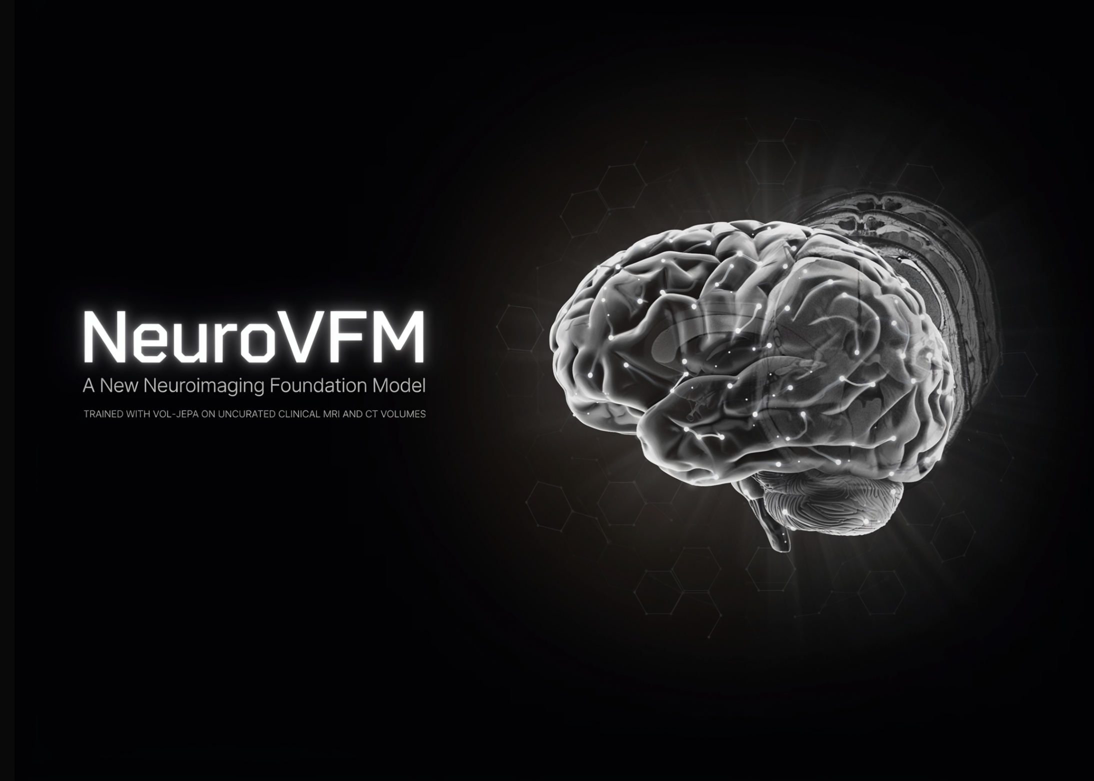
Frontier models learn mostly from public internet data. However, clinical neuroimaging rarely appears there, because MRI and CT scans contain identifiable facial features. Consequently, general models underperform on brain-imaging tasks. A University of Michigan research team addresses this gap with NeuroVFM, published in Nature Medicine.
What is NeuroVFM?
At its core, NeuroVFM is a generalist visual foundation model for neuroimaging. Specifically, it was trained on 5.24 million clinical MRI and CT volumes. These came from 566,915 studies in the UM-NeuroImages dataset. That data spans over two decades of routine care at Michigan Medicine.
The research team call their approach ‘health system learning.’ In short, the model learns from uncurated data generated during normal clinical operations. Therefore, it avoids the bottleneck of paired radiology reports. It also avoids the disease-specific curation used in narrow classifiers.
Notably, the base model is called Vol-JEPA. It extends the earlier I-JEPA and V-JEPA methods to volumetric medical images. This reflects a wider trend: JEPA-style learning is expanding into medical imaging.