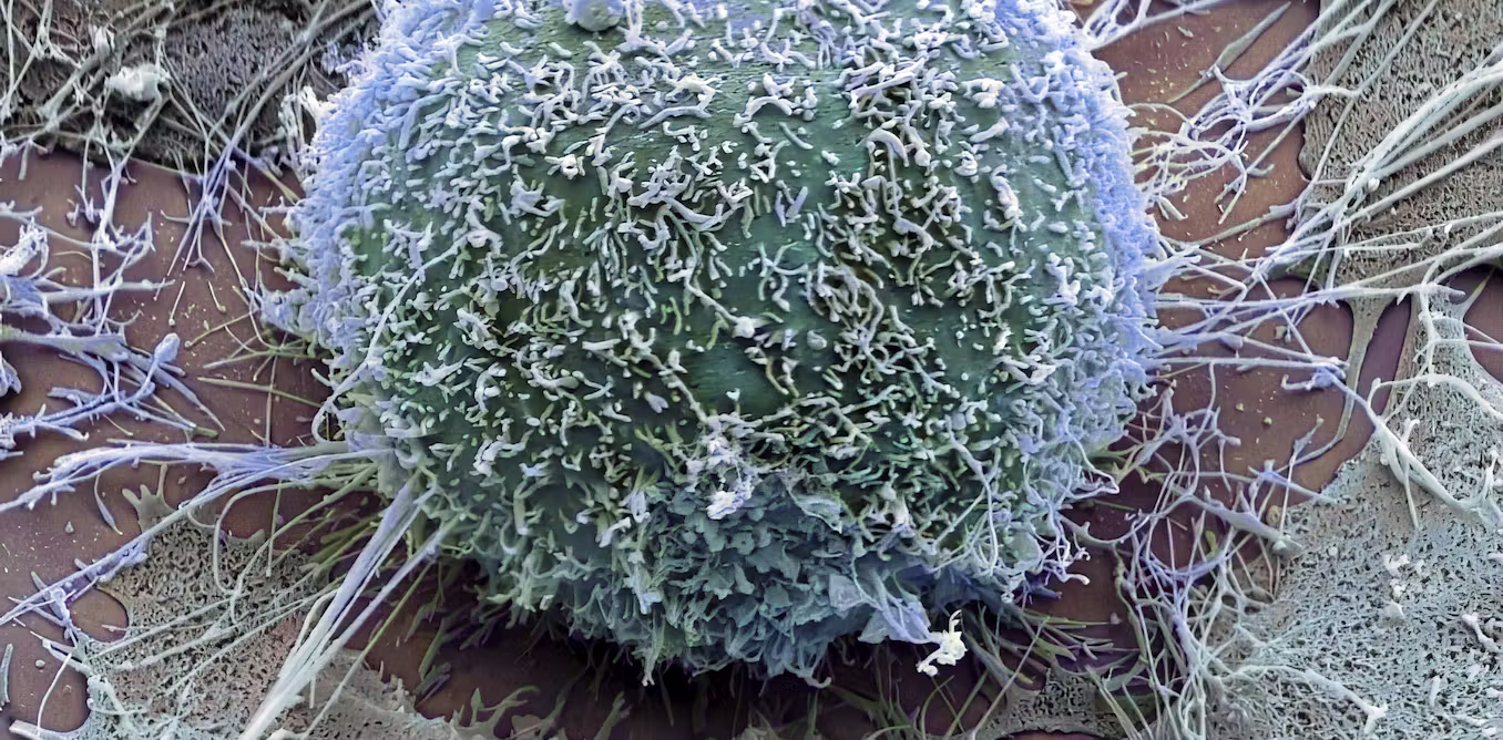
Every human cell is covered by a thin layer of sugars called the glycocalyx. This outer coating helps cells interact with their surroundings and may also provide important clues about what is happening inside the cell itself. Researchers at the Max Planck Institute for the Science of Light (MPL) have now created detailed maps of these sugar structures using advanced high resolution microscopy. Their findings, published in Nature Nanotechnology, suggest that changes in the arrangement of these sugars could one day help doctors detect diseases such as cancer.
The glycocalyx surrounds all human cells like a protective outer shell. Rather than remaining fixed in place, these complex sugar molecules constantly shift and reorganize. Scientists in the "Physical Glycosciences" research group, led by Prof. Leonhard Möckl at MPL, study how this sugar coating behaves and what it reveals about cell biology.
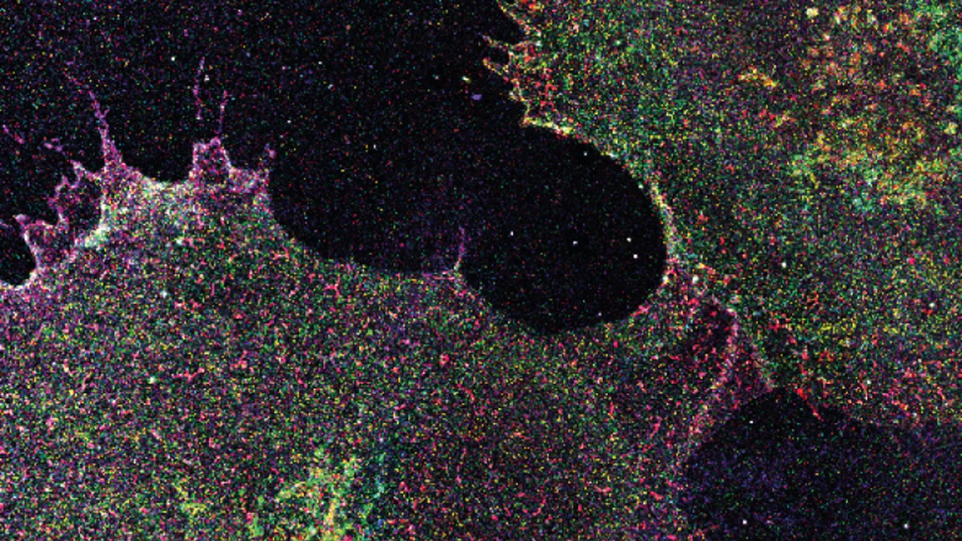
To investigate these structures, the team developed a technique called "Glycan Atlasing." Using cutting edge super resolution microscopy, they mapped the glycocalyx at the level of individual sugar molecules across many different types of cells. Their work included cell culture lines, primary human blood cells, and tissue samples.