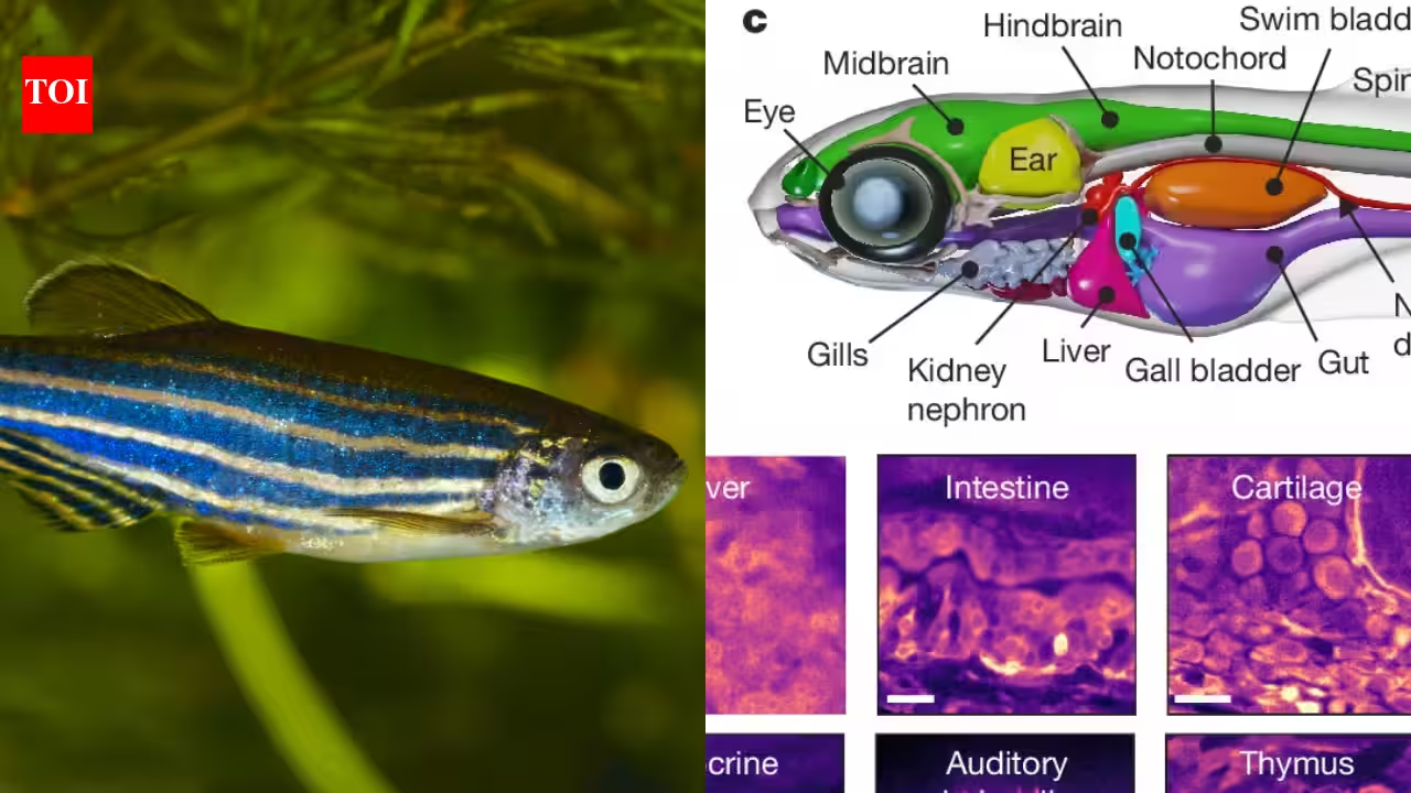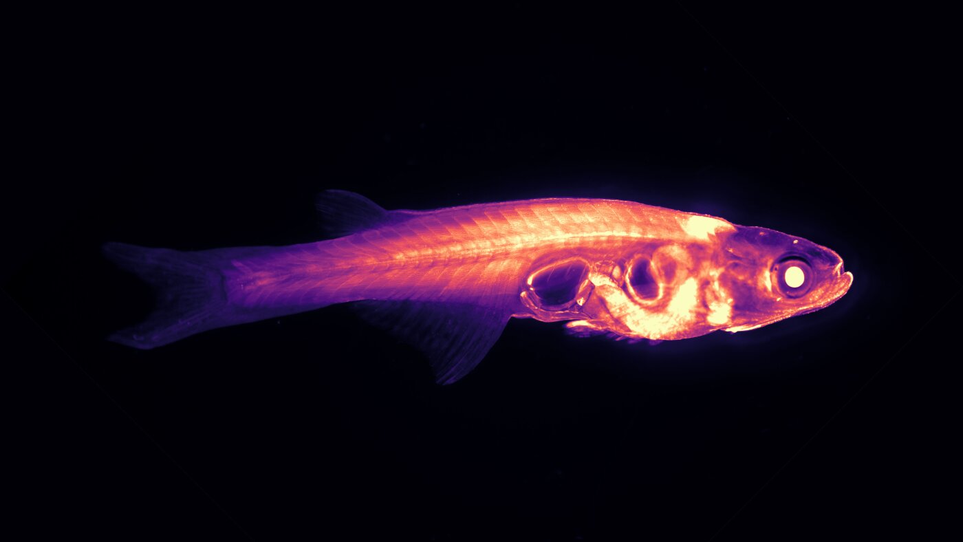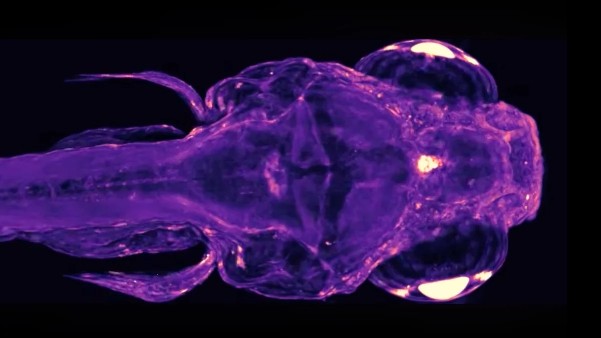
New techniques reveal previously unseen communication between living systems in the body of a zebrafish. And yes, that glowing node between its eyes is the pineal gland. (Ruetten et al., Nature, 2026)
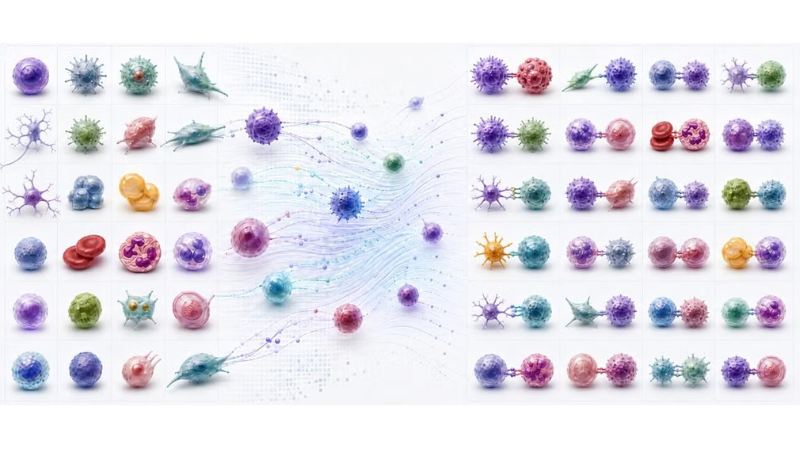
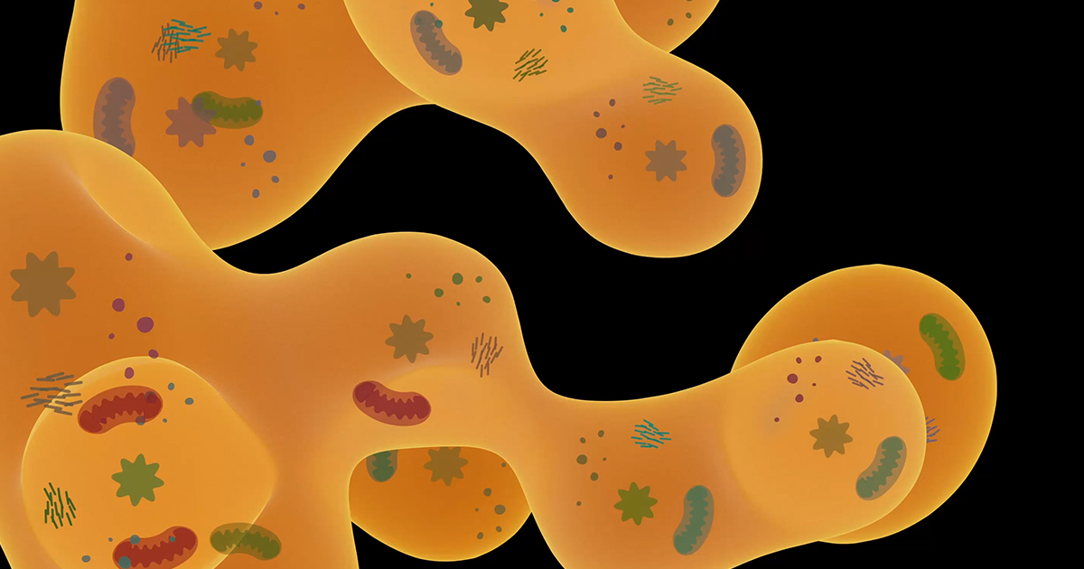
Biology – the study of living things – can admittedly be a little reductive at times.It's easy to miss important connections depending on how you look at an organism. Microscopes show us the fine details of a body, dissection has helped us identify different organs, and X-rays can show us the bigger picture of internal systems.But until now, it's been tricky to see how living cells communicate across those systems, which traditionally have been studied in isolation, even though our bodies ultimately work as a whole.Now, scientists have figured out how to watch cells from disparate parts of the body communicate with each other. Their research has been presented in the journal Nature.
"This work bridges two fundamental scales of biology – the cell and the organism – such that we can now fill that observability gap," says computational neuroscientist Virginie Ruetten, who developed the technique along with a team based at the Howard Hughes Medical Institute in the US. "There are some really basic properties that were just missing because it's been very difficult to look at cellular responses at scale."The new imaging technique – dubbed WHOLISTIC (WHole-Organism Live-Imaging System for recording Tissue and IntraCellular activity) – captures cellular activity across the entire body, second by second.So far, Ruetten and team have used this technique to image real-time signaling in naturally transparent animals: first with larval zebrafish, and then with another kind of freshwater fish, Danionella cerebrum, which remain transparent into adulthood.