A previously healthy 52-year-old woman entered the emergency department with a 4-month history of headaches and visual hallucinations. For the past month, her speech was incoherent.
When she reached the hospital, the patient was awake and alert, but unable to follow simple commands. Her speech was fluent but nonsensical, with paraphasic errors and impaired naming and repetition, reported Rolter Lorenz Lee, MD, of the University of Santo Tomas Faculty of Medicine and Surgery, and Bonifacio Pedregosa II, MD, of the Jose R. Reyes Memorial Medical Center in Manila in the Philippines, in the New England Journal of Medicine.
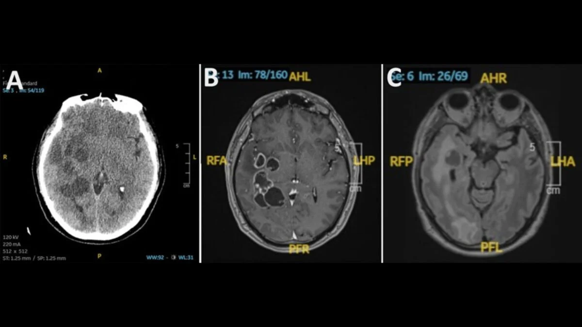
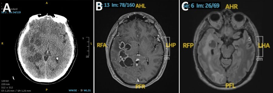
Her head CT showed several hypodense lesions with internal hyperdensities scattered throughout her brain, Lee and Pedregosa wrote. Electroencephalography (EEG) revealed posterior temporal interictal epileptiform discharges from the left side. Cerebrospinal fluid tests were normal.
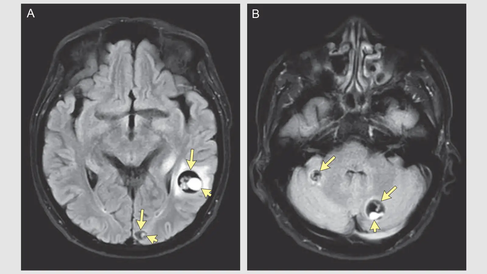
Her MRI scans showed cystic lesions in the temporal and occipital lobes on the left side, in the cerebellar hemispheres and frontal lobes bilaterally, and in the right parietal lobe. The cysts contained bright, off-center nodules representing the scolices (anterior end) of tapeworms.
"What stood out most was the exceptionally clear demonstration of the classic 'dot-in-hole' sign, a pathognomonic imaging feature of neurocysticercosis that every clinician and radiologist should recognize," Pedregosa told MedPage Today.